Login
Registration enables users to use special features of this website, such as past
order histories, retained contact details for faster checkout, review submissions, and special promotions.
order histories, retained contact details for faster checkout, review submissions, and special promotions.
Forgot password?
Registration enables users to use special features of this website, such as past
order histories, retained contact details for faster checkout, review submissions, and special promotions.
order histories, retained contact details for faster checkout, review submissions, and special promotions.
Quick Order
Products
Antibodies
ELISA and Assay Kits
Research Areas
Infectious Disease
Resources
Purchasing
Reference Material
Contact Us
Locations
Orders Processing,
Shipping & Receiving,
Warehouse
2 Shaker Rd Suites
B001/B101
Shirley, MA 01464
Production Lab
Floor 6, Suite 620
20700 44th Avenue W
Lynnwood, WA 98036
Telephone Numbers
Tel: +1 (206) 374-1102
Fax: +1 (206) 577-4565
Contact Us
Additional Contact Details
Login
Registration enables users to use special features of this website, such as past
order histories, retained contact details for faster checkout, review submissions, and special promotions.
order histories, retained contact details for faster checkout, review submissions, and special promotions.
Forgot password?
Registration enables users to use special features of this website, such as past
order histories, retained contact details for faster checkout, review submissions, and special promotions.
order histories, retained contact details for faster checkout, review submissions, and special promotions.
Quick Order
| Catalog Number | Size | Price |
|---|---|---|
| LS-C165711-200 | 200 µl (0.5 mg/ml) | $393 |
1 of 10
2 of 10
3 of 10
4 of 10
5 of 10
6 of 10
7 of 10
8 of 10
9 of 10
10 of 10
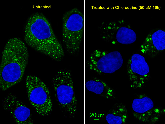
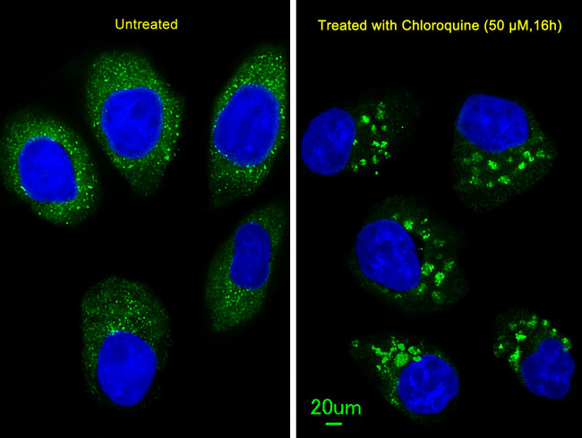
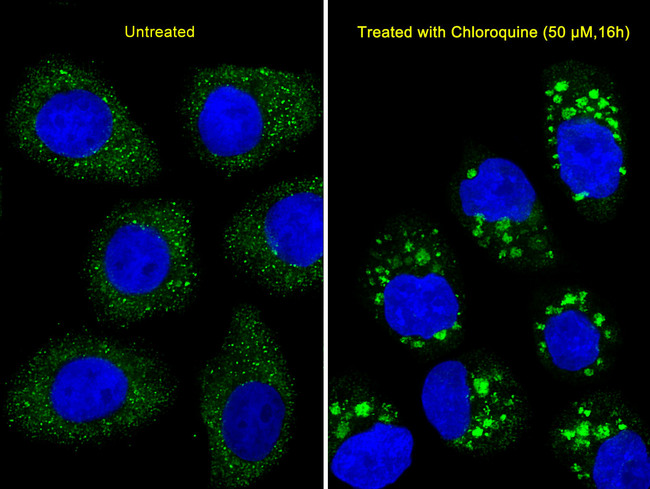
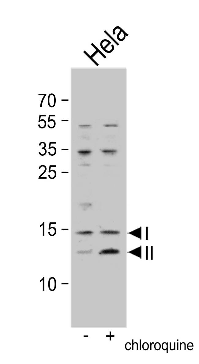
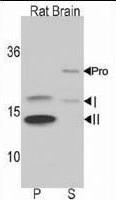
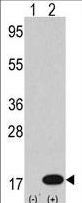
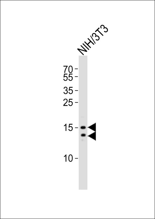
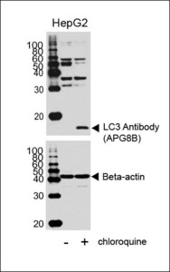
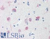
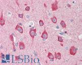
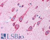
Polyclonal Rabbit anti‑Human MAP1LC3B / LC3B Antibody (aa1‑30, IHC, IF, WB) LS‑C165711
Polyclonal Rabbit anti‑Human MAP1LC3B / LC3B Antibody (aa1‑30, IHC, IF, WB) LS‑C165711

Antibody:
MAP1LC3B / LC3B Rabbit anti-Human Polyclonal (aa1-30) Antibody
Application:
IHC, IHC-P, IF, WB
Reactivity:
Human, Rat
Format:
Unconjugated, Unmodified
Toll Free North America
 206-374-1102
For Research Use Only
Overview
Antibody:
MAP1LC3B / LC3B Rabbit anti-Human Polyclonal (aa1-30) Antibody
Application:
IHC, IHC-P, IF, WB
Reactivity:
Human, Rat
Format:
Unconjugated, Unmodified
Specifications
Description
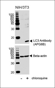
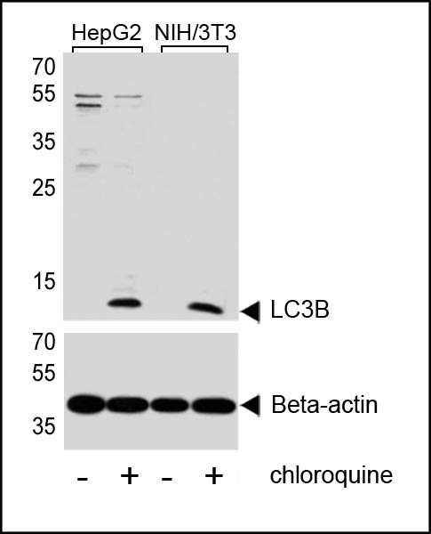
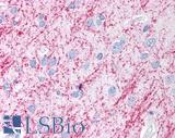
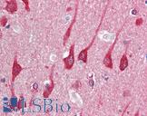
LC3B antibody LS-C165711 is an unconjugated rabbit polyclonal antibody to LC3B (MAP1LC3B) (aa1-30) from human. It is reactive with human and rat. Validated for IF, IHC and WB. Cited in 6 publications.
Target
Human MAP1LC3B / LC3B
Synonyms
MAP1LC3B | ATG8F | LC3B | MAP1A/1BLC3 | MAP1A/MAP1B LC3 B | MAP1A/MAP1B light chain 3 B | MAP1ALC3 | MAP1LC3B-a
Host
Rabbit
Reactivity
Human, Rat
(tested or 100% immunogen sequence identity)
Predicted
Cow (at least 90% immunogen sequence identity)
Clonality
Polyclonal
Conjugations
Unconjugated
Purification
Protein A purified
Modifications
Unmodified
Epitope
aa1-30
Specificity
This LC3 antibody is generated from rabbits immunized with a KLH conjugated synthetic peptide between 1-30 amino acids from the N-terminal region of human LC3.
Applications
- IHC
- IHC - Paraffin (1:50 - 1:100)
- Immunofluorescence (1:100)
- Western blot (1:1000)

|
Performing IHC? See our complete line of Immunohistochemistry Reagents including antigen retrieval solutions, blocking agents
ABC Detection Kits and polymers, biotinylated secondary antibodies, substrates and more.
|
Presentation
PBS, 0.09% Sodium Azide
Storage
Maintain refrigerated at 2°C to 8°C for up to 6 months. For long term storage store at -20°C.
Restrictions
For research use only. Intended for use by laboratory professionals.
About MAP1LC3B / LC3B
LSBio Ratings
MAP1LC3B / LC3B Antibody (aa1-30) for IHC, IF/Immunofluorescence, WB/Western LS-C165711 has an LSBio Rating of
Publications (4.5)
Learn more about The LSBio Ratings Algorithm
Publications (6)
Active ras triggers death in glioblastoma cells through hyperstimulation of macropinocytosis. Overmeyer JH, Kaul A, Johnson EE, Maltese WA. Molecular cancer research : MCR. 2008 6:965-77.
Lysosomal degradation of endocytosed proteins depends on the chloride transport protein ClC-7. Wartosch L, Fuhrmann JC, Schweizer M, Stauber T, Jentsch TJ. FASEB journal : official publication of the Federation of American Societies for Experimental Biology. 2009 23:4056-68.
Rab5 and class III phosphoinositide 3-kinase Vps34 are involved in hepatitis C virus NS4B-induced autophagy. Su WC, Chao TC, Huang YL, Weng SC, Jeng KS, Lai MM. Journal of virology. 2011 85:10561-71.
Caspase-6 activity in a BACHD mouse modulates steady-state levels of mutant huntingtin protein but is not necessary for production of a 586 amino acid proteolytic fragment. Gafni J, Papanikolaou T, Degiacomo F, Holcomb J, Chen S, Menalled L, Kudwa A, Fitzpatrick J, Miller S, Ramboz S, Tuunanen PI, Lehtimki KK, Yang XW, Park L, Kwak S, Howland D, Park H, Ellerby LM. The Journal of neuroscience : the official journal of the Society for Neuroscience. 2012 32:7454-65.
Chronic autophagy is a cellular adaptation to tumor acidic pH microenvironments. Wojtkowiak JW, Rothberg JM, Kumar V, Schramm KJ, Haller E, Proemsey JB, Lloyd MC, Sloane BF, Gillies RJ. Cancer research. 2012 72:3938-47.
Induction of autophagy by Imatinib sequesters Bcr-Abl in autophagosomes and down-regulates Bcr-Abl protein. Gafni J, Papanikolaou T, Degiacomo F, Holcomb J, Chen S, Menalled L, Kudwa A, Fitzpatrick J, Miller S, Ramboz S, Tuunanen PI, Lehtimki KK, Yang XW, Park L, Kwak S, Howland D, Park H, Ellerby LM. American journal of hematology. 2013
Customer Reviews (0)
Featured Products
Species:
Human, Mouse, Rat, Dog, Fish, Zebrafish
Applications:
IHC, IHC - Paraffin, Western blot, Immunoprecipitation
Species:
Human, Mouse, Rat, Bovine, Dog, Pig, Zebrafish, Primate
Applications:
IHC, IHC - Paraffin, IHC - Frozen, Immunofluorescence, Flow Cytometry
Species:
Human, Mouse
Applications:
IHC, IHC - Paraffin, Western blot
Species:
Human, Mouse
Applications:
IHC, IHC - Paraffin, Immunofluorescence, Western blot
Species:
Human, Rat, Zebrafish
Applications:
IHC, IHC - Paraffin, Western blot, Immunoprecipitation
Request SDS/MSDS
To request an SDS/MSDS form for this product, please contact our Technical Support department at:
Technical.Support@LSBio.com
Requested From: United States
Date Requested: 5/21/2024
Date Requested: 5/21/2024


