Login
Registration enables users to use special features of this website, such as past
order histories, retained contact details for faster checkout, review submissions, and special promotions.
order histories, retained contact details for faster checkout, review submissions, and special promotions.
Forgot password?
Registration enables users to use special features of this website, such as past
order histories, retained contact details for faster checkout, review submissions, and special promotions.
order histories, retained contact details for faster checkout, review submissions, and special promotions.
Quick Order
Products
Antibodies
ELISA and Assay Kits
Research Areas
Infectious Disease
Resources
Purchasing
Reference Material
Contact Us
Locations
Orders Processing,
Shipping & Receiving,
Warehouse
2 Shaker Rd Suites
B001/B101
Shirley, MA 01464
Production Lab
Floor 6, Suite 620
20700 44th Avenue W
Lynnwood, WA 98036
Telephone Numbers
Tel: +1 (206) 374-1102
Fax: +1 (206) 577-4565
Contact Us
Additional Contact Details
Login
Registration enables users to use special features of this website, such as past
order histories, retained contact details for faster checkout, review submissions, and special promotions.
order histories, retained contact details for faster checkout, review submissions, and special promotions.
Forgot password?
Registration enables users to use special features of this website, such as past
order histories, retained contact details for faster checkout, review submissions, and special promotions.
order histories, retained contact details for faster checkout, review submissions, and special promotions.
Quick Order
LSBio Cell-Based ELISA Kits
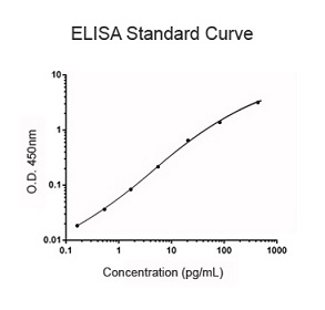
Cell-Based ELISA kits are a fast and cost-effective way to quantify specific target antigens in vitro.
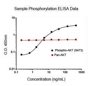
Additionally, phospho-specific cell-based ELISA kits enable researchers to quickly evaluate changes in the phosphorylation state of specific proteins in whole cells under various treatment conditions.
In a single experiment the activation state of 96 treated and untreated cells can be evaluated simultaneously.
Each kit is ready-to-use with all the necessary reagents and a clear, concise protocol that will step you through the process, from sample preparation to analysis of the results.
Cell-Based ELISA kits are available for both the detection of both phosphorylated and non-phosphorylated targets.
Features:
- Ready-to-use kit includes all necessary reagents
- Direct in vitro detection on un-lysed cultured cells
- Phospho- and non-Phospho specific kits available
- Excellent sensitivity, specificity, and reproducibility
- Built on standard 96-well microtiter format
- 450 nm colorimetric detection
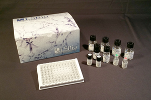
Kit Components:
- 96-well microtiter plate
- Fixing Solution
- Quenching buffer
- Blocking Buffer
- Wash Buffers
- Primary Detection Antibody
- HRP-Conjugated Secondary Antibody
- TMB Substrate Solution
- Stop Buffer
- Plate Sealer Sheets
Cell-Based ELISA Kit Protocol
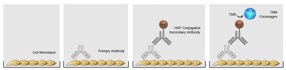
| 1) Cell monolayers are cultured in the 96-well microtiter plate. Each well can then be treated with stimulants, inhibitors, etc. The cells are then fixed and blocked. |
| 2) Antigen specific primary antibody is added and binds to the target antigen on the cells surface. |
| 3) The wells are washed and a HRP-conjugated secondary antibody is added which binds to the primary antibody. |
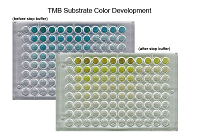
| 4) The wells are washed and a solution containing TMB is added. The TMB reacts with the HRP changing from colorless to a blue color. An acid stop solution is then added, the reaction stops, and the blue color changes to yellow. |
Phospho-Specific Cell-Based ELISA Kit Protocol
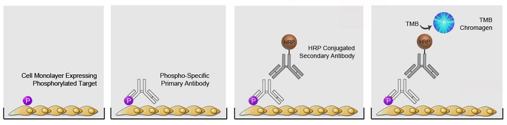
| 1) Cell monolayers are cultured in the 96-well microtiter plate. Each well can then be treated with stimulants, inhibitors, etc. to induce expression of the phosphorylated target. The cells are then fixed and blocked. |
| 2) Phospho-specific or pan-specific primary antibody is added and binds to the target antigen on the cells surface. |
| 3) The wells are washed and a HRP-conjugated secondary antibody is added which binds to the primary antibody. |
| 4) The wells are washed and a solution containing TMB is added. The TMB reacts with the HRP changing from colorless to a blue color. An acid stop solution is then added, the reaction stops, and the blue color changes to yellow. |
The plate can then be read in a spectrophotometer at a wavelength of 450 nm.