Login
Registration enables users to use special features of this website, such as past
order histories, retained contact details for faster checkout, review submissions, and special promotions.
order histories, retained contact details for faster checkout, review submissions, and special promotions.
Forgot password?
Registration enables users to use special features of this website, such as past
order histories, retained contact details for faster checkout, review submissions, and special promotions.
order histories, retained contact details for faster checkout, review submissions, and special promotions.
Quick Order
Products
Antibodies
ELISA and Assay Kits
Research Areas
Infectious Disease
Resources
Purchasing
Reference Material
Contact Us
Locations
Orders Processing,
Shipping & Receiving,
Warehouse
2 Shaker Rd Suites
B001/B101
Shirley, MA 01464
Production Lab
Floor 6, Suite 620
20700 44th Avenue W
Lynnwood, WA 98036
Telephone Numbers
Tel: +1 (206) 374-1102
Fax: +1 (206) 577-4565
Contact Us
Additional Contact Details
Login
Registration enables users to use special features of this website, such as past
order histories, retained contact details for faster checkout, review submissions, and special promotions.
order histories, retained contact details for faster checkout, review submissions, and special promotions.
Forgot password?
Registration enables users to use special features of this website, such as past
order histories, retained contact details for faster checkout, review submissions, and special promotions.
order histories, retained contact details for faster checkout, review submissions, and special promotions.
Quick Order
| Catalog Number | Size | Price |
|---|---|---|
| LS-B16379-0.1 | 0.1 mg (1 mg/ml) | $485 |
![PDCD1 / CD279 / PD-1 Antibody - Immunofluorescence of PD-1 in in overexpressing HEK293 cells with PD-1 antibody at 20 ug/mL. Green: PD1 Antibody [5D3] Blue: DAPI staining](https://lsbio-7d62.kxcdn.com/image2/pathplus-pdcd1-cd279-pd-1-antibody-clone-5d3-ls-b16379/484375_3816874.gif)
![PDCD1 / CD279 / PD-1 Antibody - Immunofluorescence of PD-1 in human lymph node tissue with PD-1 antibody at 20 ug/mL. Green: PD1 Antibody [10B3] (RF16005) Blue: DAPI staining](https://lsbio-7d62.kxcdn.com/image2/pathplus-pdcd1-cd279-pd-1-antibody-clone-5d3-ls-b16379/484374_3816821.gif)
1 of 5
2 of 5
3 of 5
4 of 5
5 of 5
PathPlus™ Monoclonal Mouse anti‑Human PDCD1 / CD279 / PD‑1 Antibody (clone 5D3, IHC, IF) LS‑B16379
PathPlus™ Monoclonal Mouse anti‑Human PDCD1 / CD279 / PD‑1 Antibody (clone 5D3, IHC, IF) LS‑B16379

Note: This antibody replaces LS-C759783
Antibody:
PDCD1 / CD279 / PD-1 Mouse anti-Human Monoclonal (5D3) Antibody
Application:
IHC-P, ICC, IF, Flo, ELISA
Reactivity:
Human
Format:
Unconjugated, Unmodified
Toll Free North America
 206-374-1102
For Research Use Only
Overview
Antibody:
PDCD1 / CD279 / PD-1 Mouse anti-Human Monoclonal (5D3) Antibody
Application:
IHC-P, ICC, IF, Flo, ELISA
Reactivity:
Human
Format:
Unconjugated, Unmodified
Specifications
Description
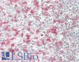
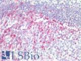
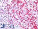
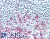
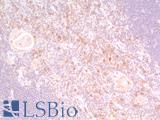
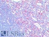
PD1 (Programmed Death Receptor 1, PDCD1, CD279) is an immune checkpoint protein active in T cells that is a target alongside its ligand PDL1 and also CTLA-4 for immunotherapy in lung and other cancers. PD1 is involved in negatively regulating T cell inflammatory activity and, when bound to receptors on tumor cells, can work to subdue tumor suppression by inhibiting the immune response. Targeted inhibition of PD1 itself can therefore function as an anti-cancer therapy by reactivating this response. PD1 is expected to have membranous staining in germinal center associated helper T cells, CD8+ T cells, and Pro-B cells. It is for the identification of subsets of T and B cell lymphomas and nodular lymphocyte predominant Hodgkin lymphomas.
References: Alsaab, 2017; Jin, 2011; Francisco, 2010; Fife, 2011; Human Pathol 2008 39(7):1050
Target
Human PDCD1 / CD279 / PD-1
Synonyms
PDCD1 | CD279 | HPD-1 | HPD-l | PD1 | Protein PD-1 | SLEB2 | PD-1 | CD279 antigen | Programmed cell death 1
Host
Mouse
Reactivity
Human
(tested or 100% immunogen sequence identity)
Clonality
IgG1
Monoclonal
Clone
5D3
Conjugations
Unconjugated
Purification
Protein A purified
Modifications
Unmodified
Immunogen
The extracellular domain of human PD-1.
Applications
- IHC - Paraffin (10 µg/ml)
- ICC
- Immunofluorescence (20 µg/ml)
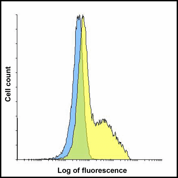
- Flow Cytometry (1 µg/ml)
- ELISA

|
Performing IHC? See our complete line of Immunohistochemistry Reagents including antigen retrieval solutions, blocking agents
ABC Detection Kits and polymers, biotinylated secondary antibodies, substrates and more.
|
Usage
Applications should be user optimized.
Presentation
PBS, 0.02% Sodium Azide, 50% Glycerol
Storage
Store at 4°C for 3 months and -20°C, stable for up to 1 year. Avoid repeated freeze-thaw cycles.
Restrictions
For research use only. Intended for use by laboratory professionals.
About PDCD1 / CD279 / PD-1
Validation
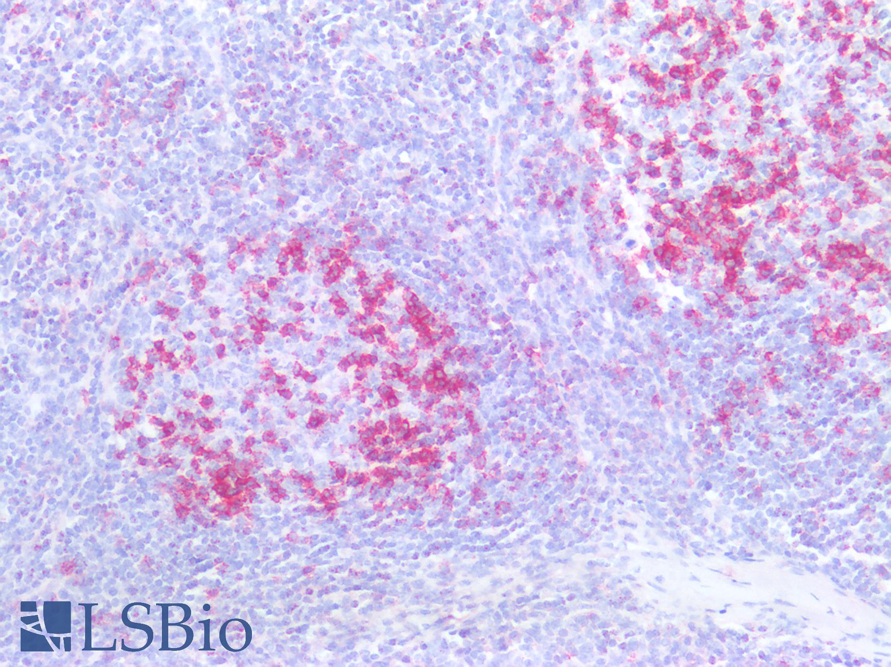
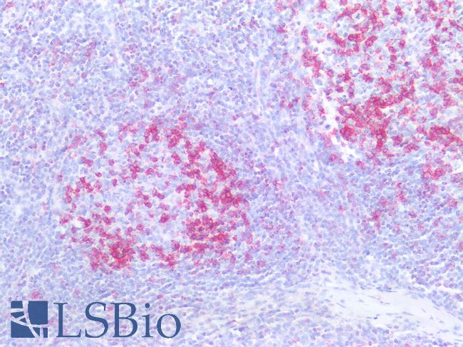
Human Tonsil: Formalin-Fixed, Paraffin-Embedded (FFPE)
Human Tonsil: Formalin-Fixed, Paraffin-Embedded (FFPE)
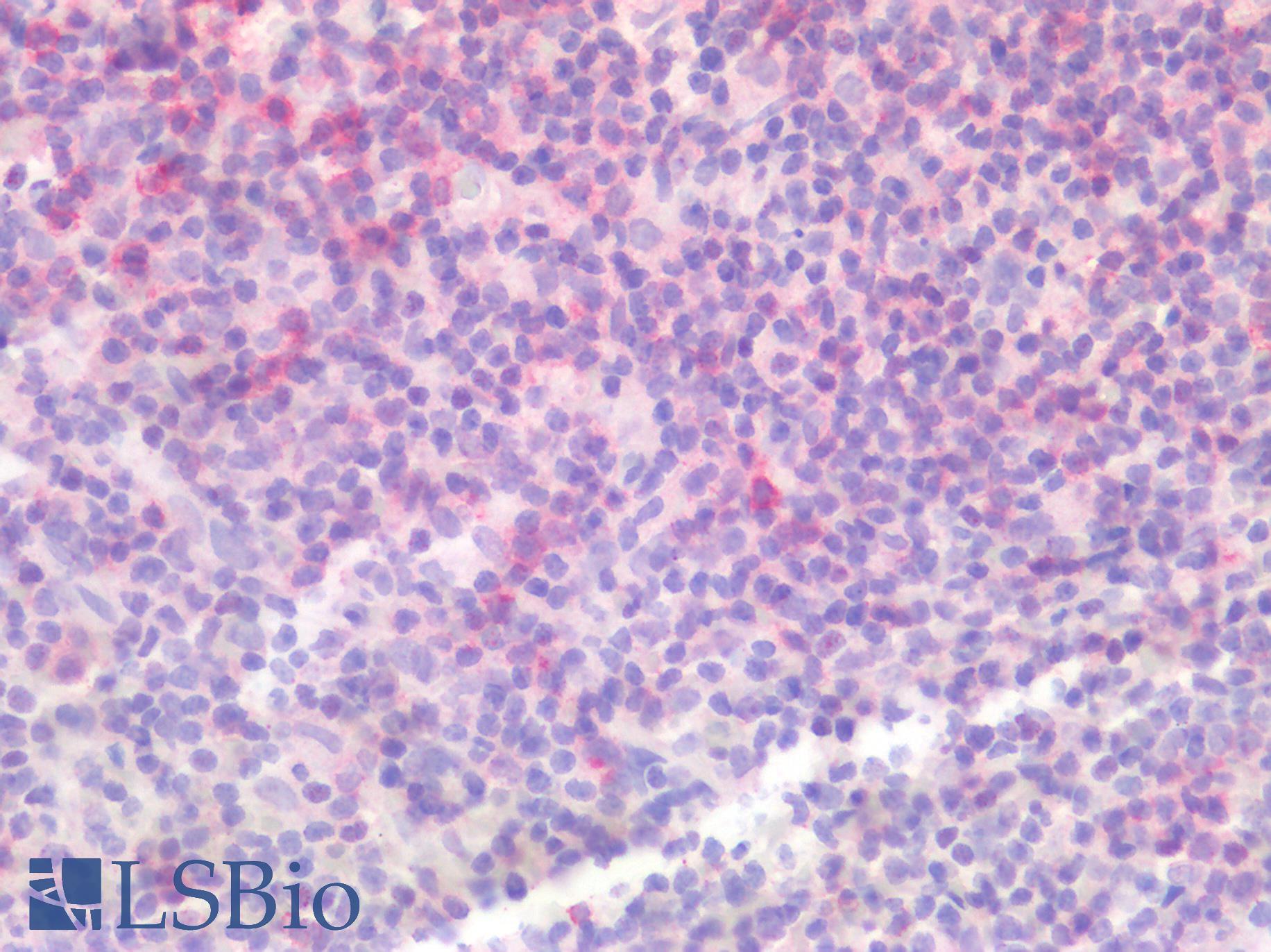
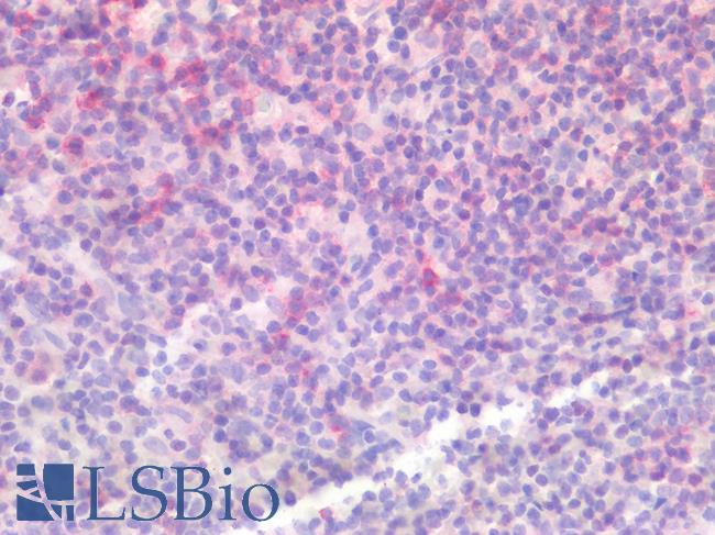
Human Spleen: Formalin-Fixed, Paraffin-Embedded (FFPE)
Human Spleen: Formalin-Fixed, Paraffin-Embedded (FFPE)
See More About...
LSBio Ratings
PathPlus™ PDCD1 / CD279 / PD-1 Antibody (clone 5D3) for IHC, ICC, IF/Immunofluorescence, Flow, ELISA LS-B16379 has an LSBio Rating of
Laboratory Validation Score (5)
Learn more about The LSBio Ratings Algorithm
Publications (0)
Customer Reviews (0)
Featured Products
Species:
Human
Applications:
IHC, IHC - Paraffin, Immunofluorescence, Western blot, Flow Cytometry
Species:
Human
Applications:
IHC, IHC - Paraffin, Flow Cytometry
Species:
Human
Applications:
IHC - Paraffin, ELISA
Species:
Human
Applications:
IHC - Paraffin, ELISA
Species:
Human, Mouse, Rat
Applications:
IHC - Paraffin, Western blot
Request SDS/MSDS
To request an SDS/MSDS form for this product, please contact our Technical Support department at:
Technical.Support@LSBio.com
Requested From: United States
Date Requested: 4/25/2024
Date Requested: 4/25/2024


