Login
Registration enables users to use special features of this website, such as past
order histories, retained contact details for faster checkout, review submissions, and special promotions.
order histories, retained contact details for faster checkout, review submissions, and special promotions.
Forgot password?
Registration enables users to use special features of this website, such as past
order histories, retained contact details for faster checkout, review submissions, and special promotions.
order histories, retained contact details for faster checkout, review submissions, and special promotions.
Quick Order
Products
Antibodies
ELISA and Assay Kits
Research Areas
Infectious Disease
Resources
Purchasing
Reference Material
Contact Us
Locations
Orders Processing,
Shipping & Receiving,
Warehouse
2 Shaker Rd Suites
B001/B101
Shirley, MA 01464
Production Lab
Floor 6, Suite 620
20700 44th Avenue W
Lynnwood, WA 98036
Telephone Numbers
Tel: +1 (206) 374-1102
Fax: +1 (206) 577-4565
Contact Us
Additional Contact Details
Login
Registration enables users to use special features of this website, such as past
order histories, retained contact details for faster checkout, review submissions, and special promotions.
order histories, retained contact details for faster checkout, review submissions, and special promotions.
Forgot password?
Registration enables users to use special features of this website, such as past
order histories, retained contact details for faster checkout, review submissions, and special promotions.
order histories, retained contact details for faster checkout, review submissions, and special promotions.
Quick Order
| Catalog Number | Size | Price |
|---|---|---|
| LS-B5565-0.1 | 0.1 ml | $460 |
1 of 2
2 of 2
PathPlus™ Monoclonal Mouse anti‑Human EPCAM Antibody (clone MOC‑31, Concentrated, IHC, WB) LS‑B5565
PathPlus™ Monoclonal Mouse anti‑Human EPCAM Antibody (clone MOC‑31, Concentrated, IHC, WB) LS‑B5565

Note: This antibody replaces LS-C134852
Antibody:
EPCAM Mouse anti-Human Monoclonal (Concentrated) (MOC-31) Antibody
Application:
IHC, IHC-P, IHC-Fr, WB
Reactivity:
Human
Format:
Unconjugated, Concentrated
Toll Free North America
 206-374-1102
For Research Use Only
Overview
Antibody:
EPCAM Mouse anti-Human Monoclonal (Concentrated) (MOC-31) Antibody
Application:
IHC, IHC-P, IHC-Fr, WB
Reactivity:
Human
Format:
Unconjugated, Concentrated
Specifications
Description
EPCAM (TACSTD1) is an epithelial membrane glycoprotein involved in cell adhesion that is present on the surface of a variety of epithelial cells. It is expressed in a wide variety of cancers, including: basal cell carcinomas, mammary Paget disease, lung adenocarcinomas, trichoepitheliomas, dermatofibromas, basal-cell carcinomas, cholangiocarcinomas, colorectal carcinomas, and carcinomas of prostate, ovary, endometrium, head and neck, and thyroid. EPCAM is useful for distinguishing epithelial cells (positive) from mesothelial cells (negative). It has also been implicated in the progression of individuals with Lynch Syndrome who hold inherited deletions in the gene. These deletions lead to silencing of the adjacent repair gene MSH2 via transcriptional read-through, which then causes microsatellite instability and colorectal cancer (as well as other malignancies that also frequently arise from Lynch Syndrome). MOC31 and BerEP4 are common monoclonal antibodies to this target.
References: JClinPathol 1990,43:213; Acta Neuropathol 1991, 83:46; Dai, 2017; Baeuerle, 2007; Kempers 2010; ModPath 2002, 15:1279
Target
Human EPCAM
Synonyms
EPCAM | 323/A3 | ACSTD1 | 17-1A | CD326 | EGP | EGP34 | Epithelial glycoprotein | GA733-2 | HNPCC8 | Ep-CAM | ESA | HEGP314 | KS 1/4 antigen | KS1/4 | KSA | M1S2 | MIC18 | MK-1 | MH99 | TROP1 | TACST-1 | TACSTD1 | CD326 antigen | CO-17A | DIAR5 | EGP-2 | EGP314 | EGP40 | Epithelial glycoprotein 314 | HEA125 | Ly74 | M4S1
Host
Mouse
Reactivity
Human
(tested or 100% immunogen sequence identity)
Clonality
IgG1
Monoclonal
Clone
MOC-31
Conjugations
Unconjugated
Purification
Tissue culture supernatant
Modifications
Concentrated
Specificity
This antibody is reactive with lung cancer associated antigens and has been studied and categorized in different clusters of reactivity patterns during the First International Workshop on Small Cell Lung Cancer Antigens held in London in April 1987. MOC-31 reacts with most epithelia, and, in lung cancer, all lung carcinomas. The membrane-associated proteins detected by MOC-31 appear to have an apparent molecular weight of 35-40 kD. MOC-31 is not reactive with normal and malignant mesothelia and therefore is especially useful for the detection of carcinoma cells in ascites or pleural effusions (4).
Applications
- IHC
- IHC - Paraffin (1:100)
- IHC - Frozen
- Western blot

|
Performing IHC? See our complete line of Immunohistochemistry Reagents including antigen retrieval solutions, blocking agents
ABC Detection Kits and polymers, biotinylated secondary antibodies, substrates and more.
|
Usage
The antibody is useful in immunohistochemistry and immunoblotting. MOC-31 react with antigens detectable in cryostat section. For use on frozen tissue and paraffin tissue after antigen retrieval. Using the antigen retrieval techniques this antibody discriminates between cells which originate from mesothelium and epithelium. General procedure to perform cryostat sectioning and immunostaining: Cryostat sectioning and immunostaining is done as described by de Leij et al. Sections (about 6 micron thick) are cut in a cryostat at -20°C and placed on glass slides. After drying at RT and fixation in acetone (water-free), the sections are washed 2-3 times in PBS. Subsequently (after drying the glass area around the sections) 25 ul of undiluted monoclonal antibody preparation is applied to the wet sections. After incubation for 45 mins. in a humidified atmosphere, the sections are washed again in PBS (2-3 times) and the second step reagent (an appropriately diluted HRPO-conjugated, anti mouse Ig preparation, supplemented with 1% human serum) is applied to the wet sections. After incubation for an additional 20 mins., the sections are washed again in PBS (2-3) times and the staining reaction is performed with 3-aminoethylcarbazole (10 mg dissolved in 2.5 ml dimethylformamide, subsequently 0.05 M acetate buffer, pH 4.9, is added to a total volume of 50 ml, after which the solution is filtrated and H2O2 is added to a final concentration of about 0.03%). A positive reaction is indicated by red deposit. The nuclei of the cells present in the sections are counterstained with Mayer's hematoxylin to obtain a good histological picture. Working dilution: approx 1:20.
Presentation
Tissue culture supernatant (8% FBS), <0.1% sodium azide
Storage
Store at 2-8°C.
Restrictions
For research use only. Intended for use by laboratory professionals.
About EPCAM
Validation
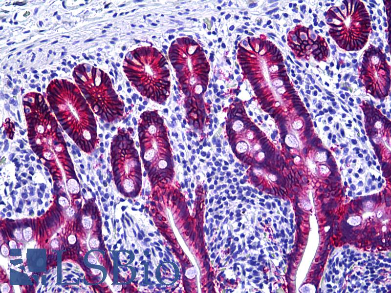
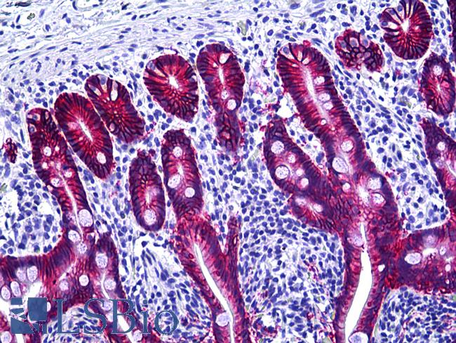
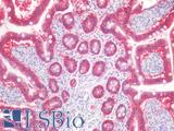
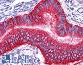
Anti-EPCAM antibody IHC of human intestine. Immunohistochemistry of formalin-fixed, paraffin-embedded tissue after heat-induced antigen retrieval. Antibody dilution 1:100.
Anti-EPCAM antibody IHC of human intestine. Immunohistochemistry of formalin-fixed, paraffin-embedded tissue after heat-induced antigen retrieval. Antibody dilution 1:100.
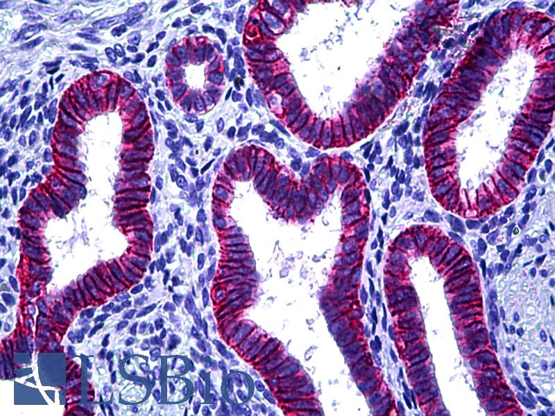
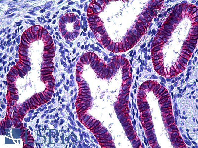
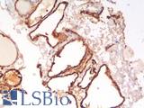
Anti-EPCAM antibody IHC of human uterus, endometrium. Immunohistochemistry of formalin-fixed, paraffin-embedded tissue after heat-induced antigen retrieval. Antibody dilution 1:100.
Anti-EPCAM antibody IHC of human uterus, endometrium. Immunohistochemistry of formalin-fixed, paraffin-embedded tissue after heat-induced antigen retrieval. Antibody dilution 1:100.
See More About...
LSBio Ratings
PathPlus™ EPCAM Antibody (clone MOC-31, Concentrated) for IHC, WB/Western LS-B5565 has an LSBio Rating of
Laboratory Validation Score (5)
Learn more about The LSBio Ratings Algorithm
Publications (0)
Customer Reviews (0)
Featured Products
Species:
Human
Applications:
IHC - Paraffin, Immunofluorescence, Flow Cytometry
Species:
Human
Applications:
IHC, IHC - Paraffin, IHC - Frozen, ICC, Western blot, Immunoprecipitation, Flow Cytometry
Species:
Mouse
Applications:
IHC, IHC - Frozen, ICC, Western blot, Immunoprecipitation, Flow Cytometry
Request SDS/MSDS
To request an SDS/MSDS form for this product, please contact our Technical Support department at:
Technical.Support@LSBio.com
Requested From: United States
Date Requested: 4/25/2024
Date Requested: 4/25/2024


