Login
Registration enables users to use special features of this website, such as past
order histories, retained contact details for faster checkout, review submissions, and special promotions.
order histories, retained contact details for faster checkout, review submissions, and special promotions.
Forgot password?
Registration enables users to use special features of this website, such as past
order histories, retained contact details for faster checkout, review submissions, and special promotions.
order histories, retained contact details for faster checkout, review submissions, and special promotions.
Quick Order
Products
Antibodies
ELISA and Assay Kits
Research Areas
Infectious Disease
Resources
Purchasing
Reference Material
Contact Us
Locations
Orders Processing,
Shipping & Receiving,
Warehouse
2 Shaker Rd Suites
B001/B101
Shirley, MA 01464
Production Lab
Floor 6, Suite 620
20700 44th Avenue W
Lynnwood, WA 98036
Telephone Numbers
Tel: +1 (206) 374-1102
Fax: +1 (206) 577-4565
Contact Us
Additional Contact Details
Login
Registration enables users to use special features of this website, such as past
order histories, retained contact details for faster checkout, review submissions, and special promotions.
order histories, retained contact details for faster checkout, review submissions, and special promotions.
Forgot password?
Registration enables users to use special features of this website, such as past
order histories, retained contact details for faster checkout, review submissions, and special promotions.
order histories, retained contact details for faster checkout, review submissions, and special promotions.
Quick Order
| Catalog Number | Size | Price |
|---|---|---|
| LS-B3862-50 | 50 µl | $440 |
1 of 8
2 of 8
3 of 8
4 of 8
5 of 8
6 of 8
7 of 8
8 of 8
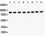
PathPlus™ Monoclonal Mouse anti‑Human BRAF / B‑Raf Antibody (clone 1H12, IHC, IF, WB) LS‑B3862
PathPlus™ Monoclonal Mouse anti‑Human BRAF / B‑Raf Antibody (clone 1H12, IHC, IF, WB) LS‑B3862

Note: This antibody replaces LS-C108748, LS-C758759, LS-C355318
Antibody:
BRAF / B-Raf Mouse anti-Human Monoclonal (1H12) Antibody
Application:
IHC-P, IF, WB, ELISA
Reactivity:
Human
Format:
Unconjugated, Unmodified
Toll Free North America
 206-374-1102
For Research Use Only
Overview
Antibody:
BRAF / B-Raf Mouse anti-Human Monoclonal (1H12) Antibody
Application:
IHC-P, IF, WB, ELISA
Reactivity:
Human
Format:
Unconjugated, Unmodified
Specifications
Description
BRAF is a MAPK/ERK signaling pathway kinase involved in regulating growth, proliferation, apoptosis and survival of cells. It is one of the most frequently somatically mutated genes in cancer, particularly in melanomas, thyroid carcinomas, and colorectal cancer. BRAF mutations (most frequently the V600E mutation) occur in up to 60% of melanomas and more than 60% of papillary thyroid carcinomas. BRAF mutation is also present in roughly 10% of colorectal cancers, where mutation detection is useful for differentiating CIMP (CpG Island Methylator Phenotype, BRAF V600E positive) from Lynch syndrome (typically BRAF mutation negative). Status of BRAF mutation will predict response to therapy in cases of advanced melanoma. Furthermore, BRAF-mutant tumors tend to be more aggressive and are linked with shorter survival. Thus, determining presence of BRAF mutation is an important step in the differential diagnosis of numerous malignancies. Staining of wild-type BRAF may be nuclear, cytoplasmic or membranous and is present in all tissues.
References: Nature Biotechnology. 2016. 34 (2):155–63, PMID: 26619011; Endocr Relat Cancer. 2014 Jan 30;21(2):161-73, PMID: 24243688; Mod Pathol. 2018 Jan; 31(1): 24–38, PMID: 29148538; Am J Transl Res. 2018; 10(8): 2726–2736, PMID: 30210710; Molecular Cancer Therapeutics. 2011 Mar. 10 (3):385, DOI: 10.1158/1535-7163.MCT-10-0799
Target
Human BRAF / B-Raf
Synonyms
BRAF | 94 kDa B-raf protein | B-raf | BRAF1 | B Raf | Proto-oncogene B-Raf | p94 | B-RAF1 | NS7 | ONCOGENE BRAF | RAFB1
Host
Mouse
Reactivity
Human
(tested or 100% immunogen sequence identity)
Clonality
IgG1
Monoclonal
Clone
1H12
Conjugations
Unconjugated
Purification
Ascites
Modifications
Unmodified
Immunogen
Purified recombinant fragment of human BRAF expressed in E. Coli encoding aa 285-401.
Specificity
Human, Mouse
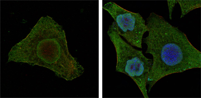
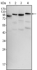
Applications
- IHC - Paraffin (1:200 - 1:400)
- Immunofluorescence (1:200 - 1:1000)
- Western blot (1:100 - 1:500)
- ELISA (1:10000)

|
Performing IHC? See our complete line of Immunohistochemistry Reagents including antigen retrieval solutions, blocking agents
ABC Detection Kits and polymers, biotinylated secondary antibodies, substrates and more.
|
Presentation
Ascitic fluid, 0.03% sodium azide
Storage
Short term: store at 4°C. Long term: store at -20°C.
Restrictions
For research use only. Intended for use by laboratory professionals.
About BRAF / B-Raf
Validation
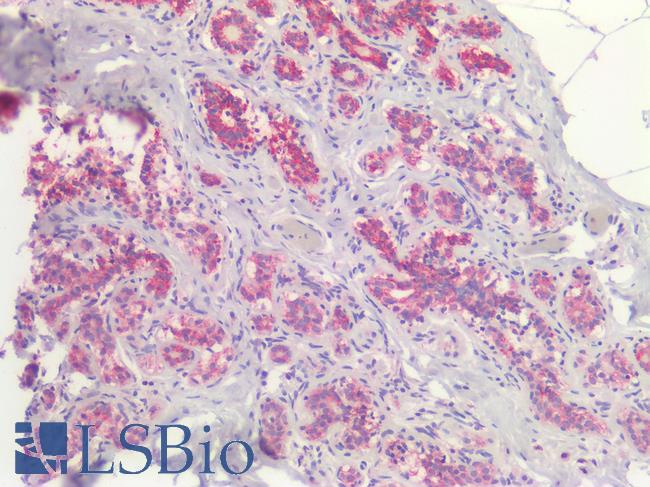
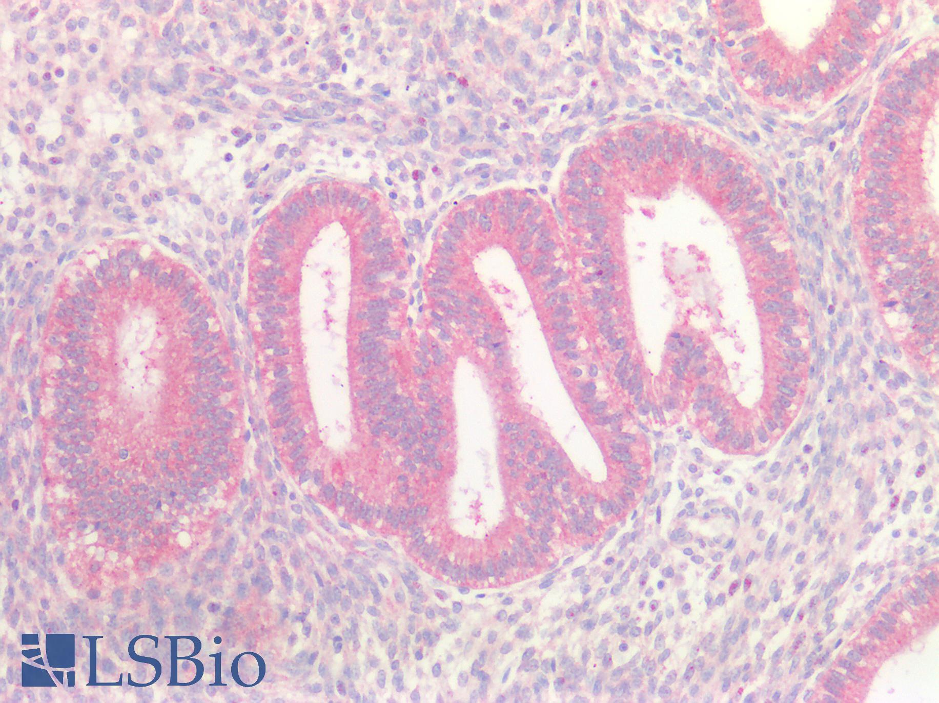
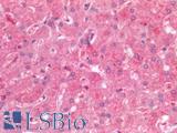
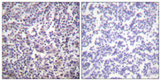
Human Breast, Carcinoma: Formalin-Fixed, Paraffin-Embedded (FFPE)
Human Breast, Carcinoma: Formalin-Fixed, Paraffin-Embedded (FFPE)
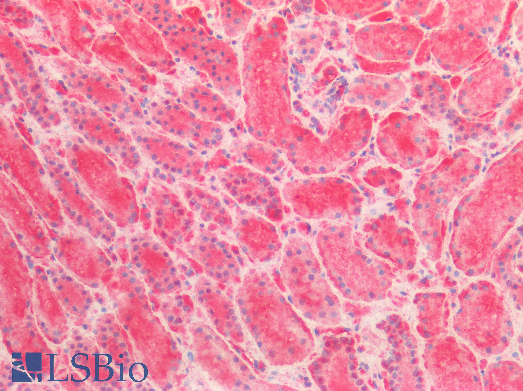
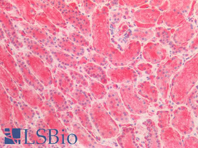
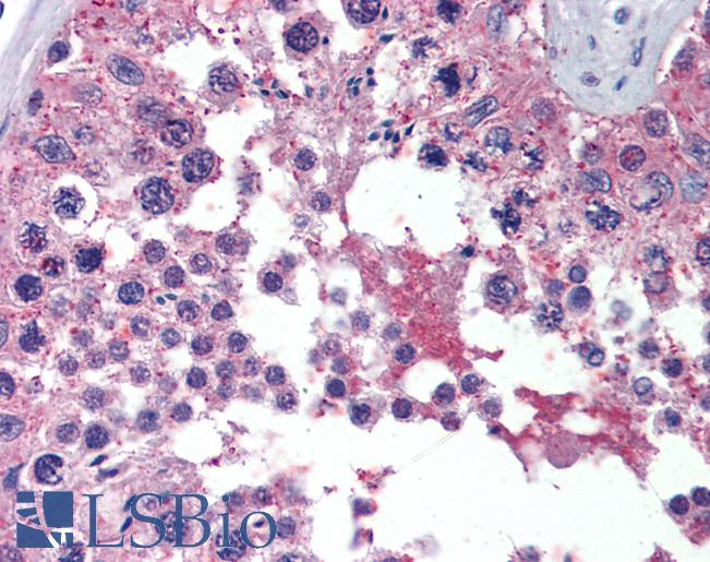
Human Kidney: Formalin-Fixed, Paraffin-Embedded (FFPE)
Human Kidney: Formalin-Fixed, Paraffin-Embedded (FFPE)
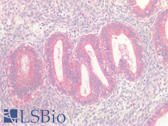
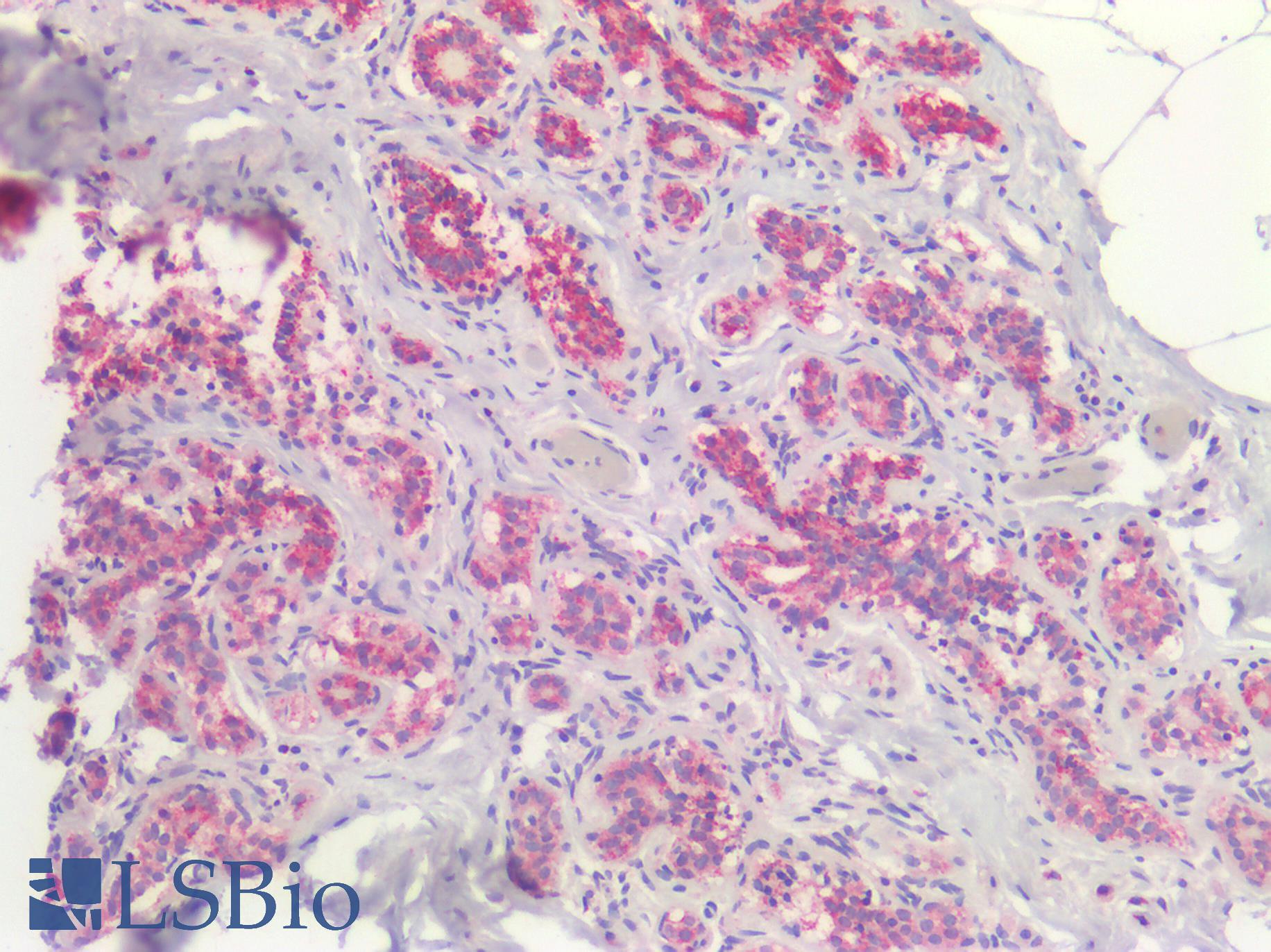
Human Uterus: Formalin-Fixed, Paraffin-Embedded (FFPE)
Human Uterus: Formalin-Fixed, Paraffin-Embedded (FFPE)
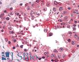
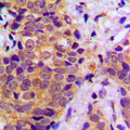
Anti-BRAF antibody IHC of human brain, cerebellum. Immunohistochemistry of formalin-fixed, paraffin-embedded tissue after heat-induced antigen retrieval. Antibody dilution 1:200.
Anti-BRAF antibody IHC of human brain, cerebellum. Immunohistochemistry of formalin-fixed, paraffin-embedded tissue after heat-induced antigen retrieval. Antibody dilution 1:200.

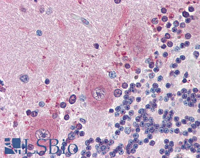
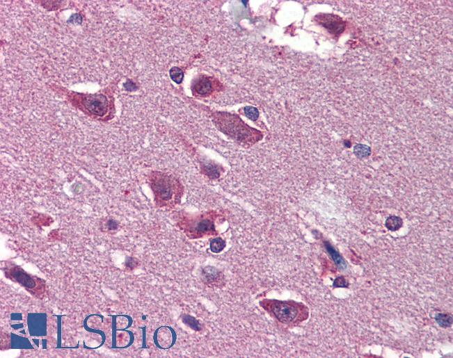
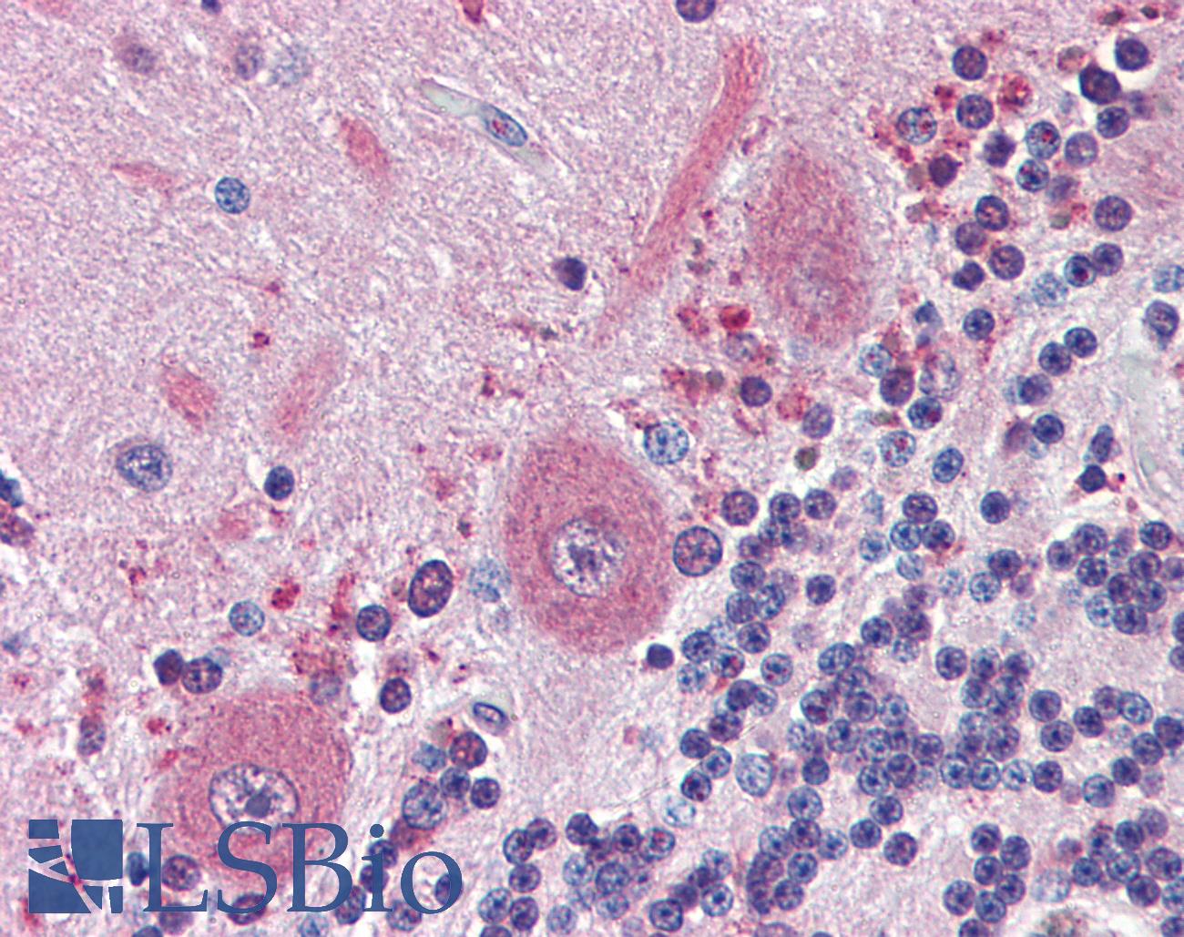
Anti-BRAF antibody IHC of human brain, cortex. Immunohistochemistry of formalin-fixed, paraffin-embedded tissue after heat-induced antigen retrieval. Antibody dilution 1:200.
Anti-BRAF antibody IHC of human brain, cortex. Immunohistochemistry of formalin-fixed, paraffin-embedded tissue after heat-induced antigen retrieval. Antibody dilution 1:200.
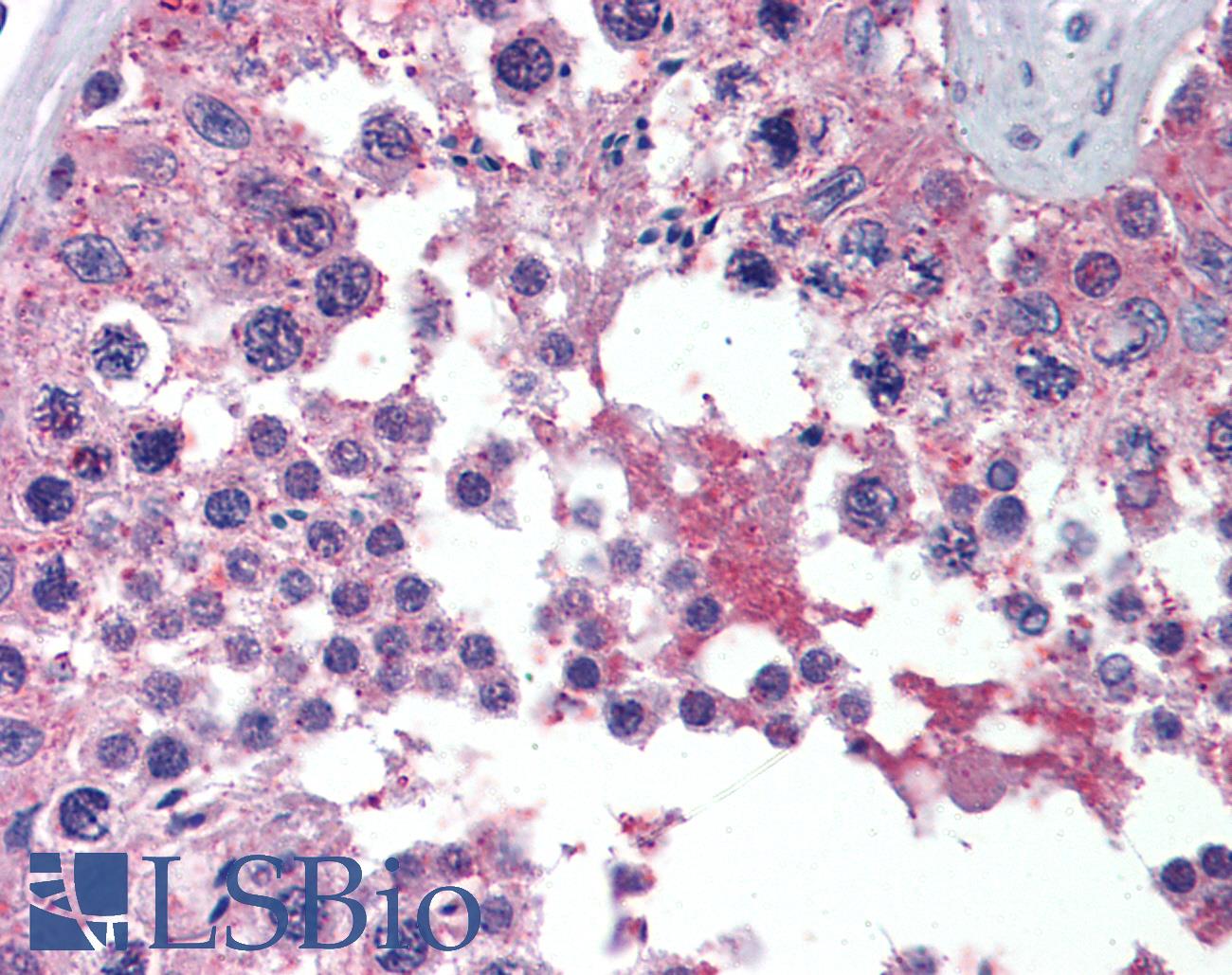
Anti-BRAF antibody IHC of human testis. Immunohistochemistry of formalin-fixed, paraffin-embedded tissue after heat-induced antigen retrieval. Antibody dilution 1:200.
Anti-BRAF antibody IHC of human testis. Immunohistochemistry of formalin-fixed, paraffin-embedded tissue after heat-induced antigen retrieval. Antibody dilution 1:200.
See More About...
LSBio Ratings
PathPlus™ BRAF / B-Raf Antibody (clone 1H12) for IHC, IF/Immunofluorescence, WB/Western, ELISA LS-B3862 has an LSBio Rating of
Laboratory Validation Score (5)
Learn more about The LSBio Ratings Algorithm
Publications (0)
Customer Reviews (0)
Featured Products
Species:
Human, Mouse, Rat
Applications:
IHC - Paraffin, Immunofluorescence, Western blot, ELISA
Species:
Human, Mouse, Rat
Applications:
IHC, Western blot, Peptide Enzyme-Linked Immunosorbent Assay
Species:
Human
Applications:
IHC, IHC - Paraffin, Western blot, ELISA
Species:
Human, Mouse, Rat, Chicken, Zebrafish
Applications:
IHC, IHC - Paraffin, Western blot
Request SDS/MSDS
To request an SDS/MSDS form for this product, please contact our Technical Support department at:
Technical.Support@LSBio.com
Requested From: United States
Date Requested: 4/26/2024
Date Requested: 4/26/2024


