Login
Registration enables users to use special features of this website, such as past
order histories, retained contact details for faster checkout, review submissions, and special promotions.
order histories, retained contact details for faster checkout, review submissions, and special promotions.
Forgot password?
Registration enables users to use special features of this website, such as past
order histories, retained contact details for faster checkout, review submissions, and special promotions.
order histories, retained contact details for faster checkout, review submissions, and special promotions.
Quick Order
Products
Antibodies
ELISA and Assay Kits
Research Areas
Infectious Disease
Resources
Purchasing
Reference Material
Contact Us
Locations
Orders Processing,
Shipping & Receiving,
Warehouse
2 Shaker Rd Suites
B001/B101
Shirley, MA 01464
Production Lab
Floor 6, Suite 620
20700 44th Avenue W
Lynnwood, WA 98036
Telephone Numbers
Tel: +1 (206) 374-1102
Fax: +1 (206) 577-4565
Contact Us
Additional Contact Details
Login
Registration enables users to use special features of this website, such as past
order histories, retained contact details for faster checkout, review submissions, and special promotions.
order histories, retained contact details for faster checkout, review submissions, and special promotions.
Forgot password?
Registration enables users to use special features of this website, such as past
order histories, retained contact details for faster checkout, review submissions, and special promotions.
order histories, retained contact details for faster checkout, review submissions, and special promotions.
Quick Order
| Catalog Number | Size | Price |
|---|---|---|
| LS-C801116-100 | 100 µl (1 mg/ml) | $379 |
| LS-C801116-200 | 200 µl (1 mg/ml) | $421 |

1 of 42
2 of 42
3 of 42
4 of 42
5 of 42
6 of 42
7 of 42
8 of 42
9 of 42
10 of 42
11 of 42
12 of 42
13 of 42
14 of 42
15 of 42
16 of 42
17 of 42
18 of 42
19 of 42
20 of 42
21 of 42
22 of 42
23 of 42
24 of 42
25 of 42
26 of 42
27 of 42
28 of 42
29 of 42
30 of 42
31 of 42
32 of 42
33 of 42
34 of 42
35 of 42
36 of 42
37 of 42
38 of 42
39 of 42
40 of 42
41 of 42
42 of 42
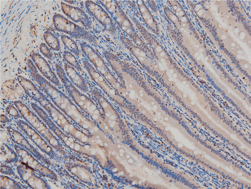
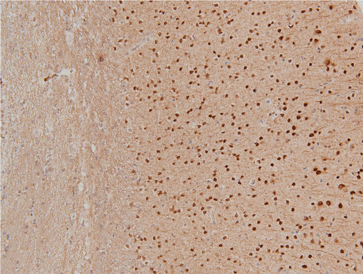
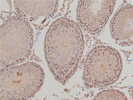
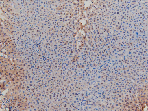
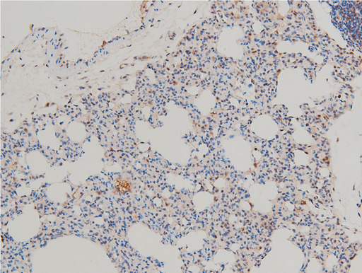
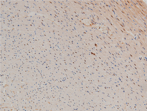
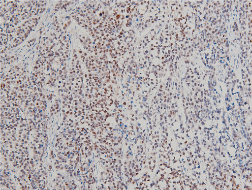
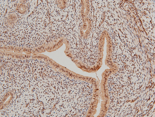
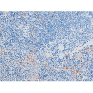
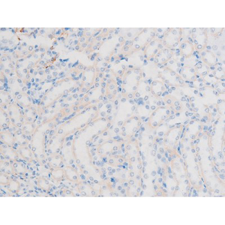
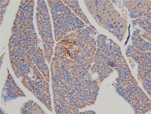
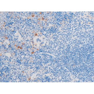
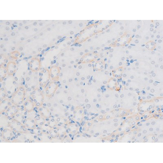
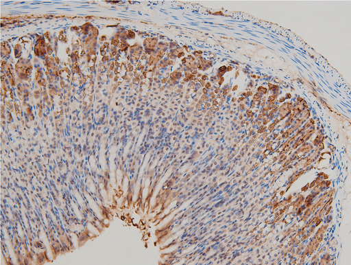
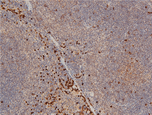
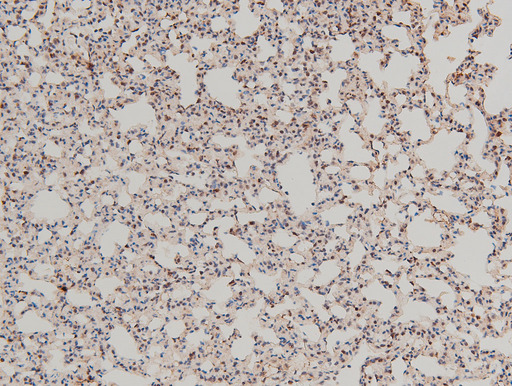
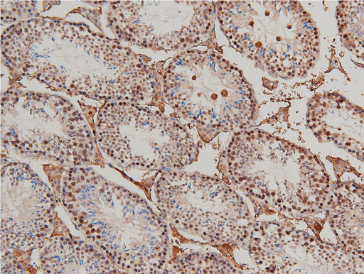
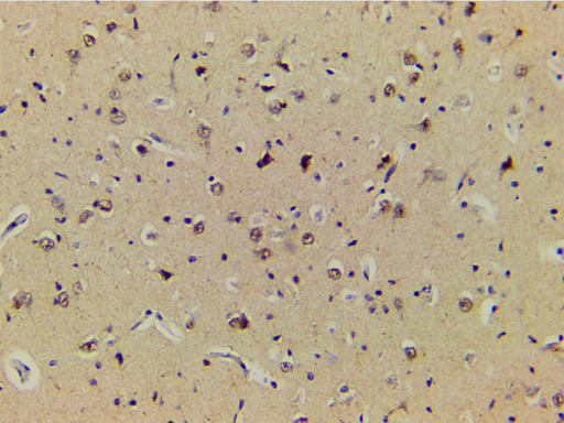
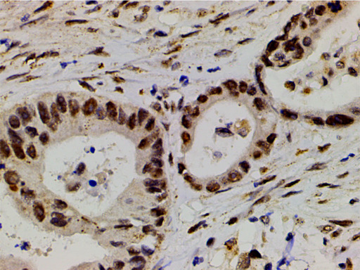
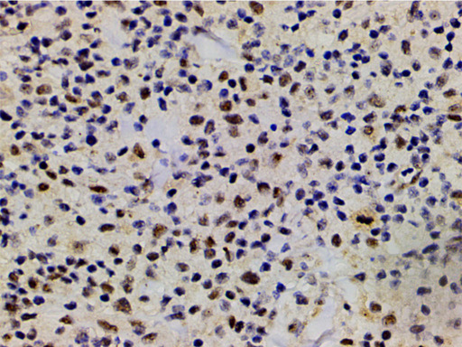
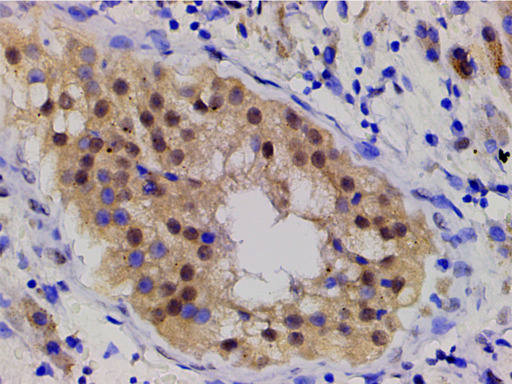
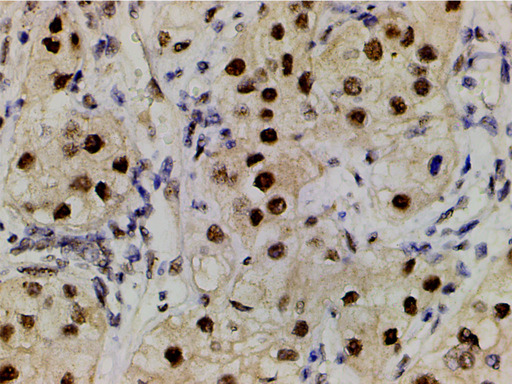
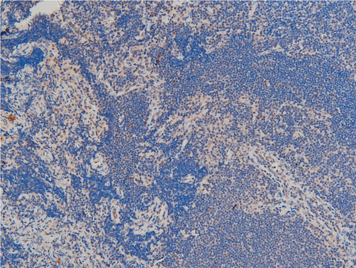
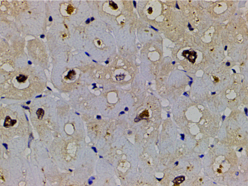
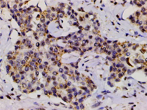
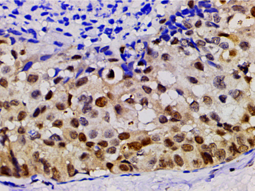
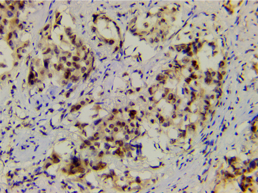
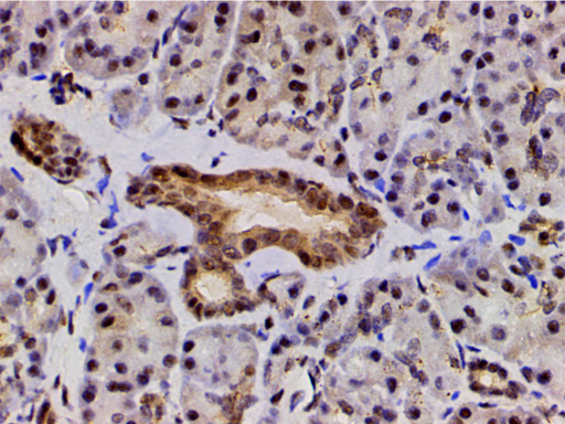
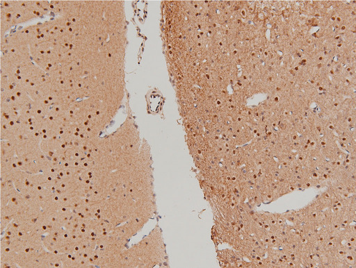
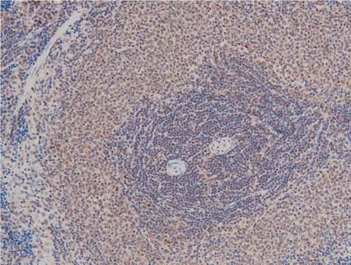
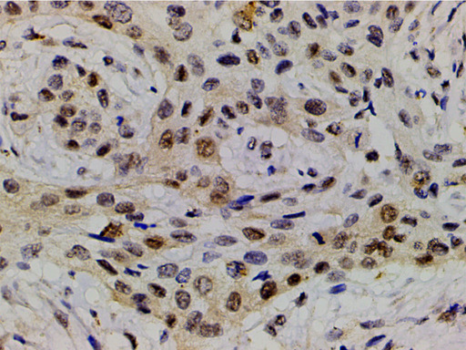
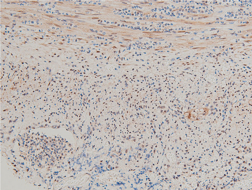
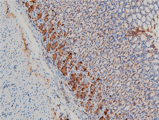
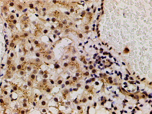
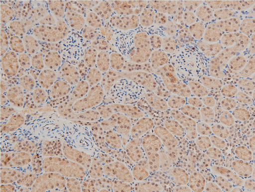
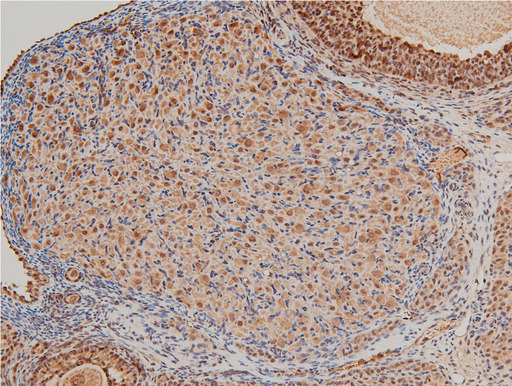
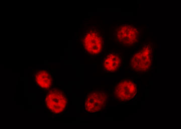
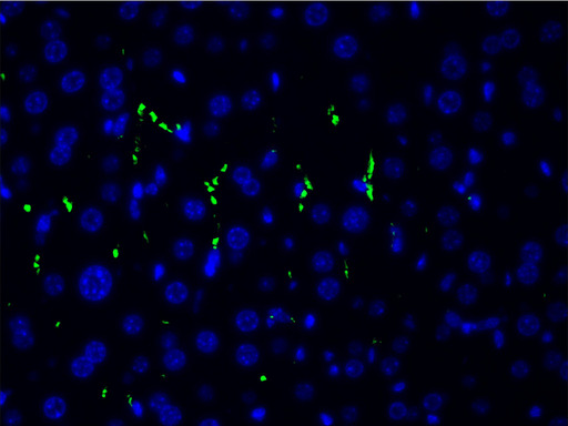
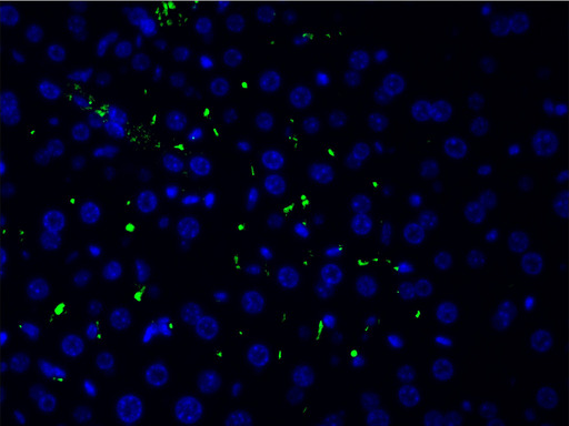
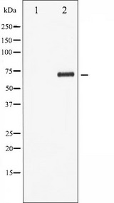
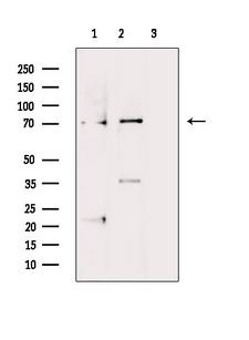
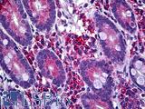
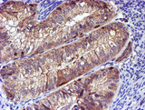
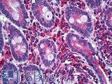
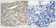
Polyclonal Rabbit anti‑Human LIMK1 / LIMK Antibody (phospho‑Thr508, IHC, IF, WB) LS‑C801116
Polyclonal Rabbit anti‑Human LIMK1 / LIMK Antibody (phospho‑Thr508, IHC, IF, WB) LS‑C801116
Antibody:
LIMK1 / LIMK Rabbit anti-Human Polyclonal (pThr508) Antibody
Application:
IHC, IF, WB, Peptide-ELISA
Reactivity:
Human, Mouse, Rat
Format:
Unconjugated, Unmodified
Toll Free North America
 206-374-1102
For Research Use Only
Overview
Antibody:
LIMK1 / LIMK Rabbit anti-Human Polyclonal (pThr508) Antibody
Application:
IHC, IF, WB, Peptide-ELISA
Reactivity:
Human, Mouse, Rat
Format:
Unconjugated, Unmodified
Specifications
Description
LIMK antibody LS-C801116 is an unconjugated rabbit polyclonal antibody to LIMK (LIMK1) (pThr508) from human. It is reactive with human, mouse and rat. Validated for IF, IHC, Peptide-ELISA and WB.
Target
Human LIMK1 / LIMK
Synonyms
LIMK1 | LIM domain kinase 1 | Lim kinase 1 | LIMK-1 | LIM kinase | LIMK
Host
Rabbit
Reactivity
Human, Mouse, Rat
(tested or 100% immunogen sequence identity)
Clonality
IgG
Polyclonal
Conjugations
Unconjugated
Purification
Affinity purification via sequential chromatography on phospho- and non-phospho-peptide affinity columns.
Modifications
Unmodified
Immunogen
A synthesized peptide derived from human LIMK1 around the phosphorylation site of Threonine 508.
Epitope
pThr508
Specificity
Phospho-LIMK1 (Thr508) Antibody detects endogenous levels of LIMK1 only when phosphorylated at Threonine 508.
Applications
- IHC (1:50 - 1:200)
- Immunofluorescence (1:100 - 1:500)
- Western blot (1:500 - 1:2000)
- Peptide Enzyme-Linked Immunosorbent Assay (1:20000 - 1:40000)

|
Performing IHC? See our complete line of Immunohistochemistry Reagents including antigen retrieval solutions, blocking agents
ABC Detection Kits and polymers, biotinylated secondary antibodies, substrates and more.
|
Usage
For western blots: incubate membrane with diluted antibody overnight in 5% w/v milk , 1X TBS, 0.1% Tween-20 at 4°C with gentle shaking.
Presentation
PBS, pH 7.4, 0.02% Sodium Azide, 50% Glycerol
Storage
Upon receipt, store at -20°C. Avoid freeze-thaw cycles.
Restrictions
For research use only. Intended for use by laboratory professionals.
About LIMK1 / LIMK
Publications (0)
Customer Reviews (0)
Featured Products
Species:
Human, Mouse, Rat
Applications:
IHC, IHC - Paraffin, ELISA
Species:
Human, Monkey, Mouse, Rat, Dog
Applications:
IHC, IHC - Paraffin, Immunofluorescence, Western blot, Flow Cytometry
Species:
Mouse
Applications:
Western blot, ELISA
Species:
Human
Applications:
IHC, IHC - Paraffin, Western blot, ELISA
Species:
Human, Mouse, Rat
Applications:
IHC, Western blot, ELISA
Reactivity:
Human, Mouse, Rat
Range:
Positive/Negative
Request SDS/MSDS
To request an SDS/MSDS form for this product, please contact our Technical Support department at:
Technical.Support@LSBio.com
Requested From: United States
Date Requested: 4/28/2024
Date Requested: 4/28/2024


