Login
Registration enables users to use special features of this website, such as past
order histories, retained contact details for faster checkout, review submissions, and special promotions.
order histories, retained contact details for faster checkout, review submissions, and special promotions.
Forgot password?
Registration enables users to use special features of this website, such as past
order histories, retained contact details for faster checkout, review submissions, and special promotions.
order histories, retained contact details for faster checkout, review submissions, and special promotions.
Quick Order
Products
Antibodies
ELISA and Assay Kits
Research Areas
Infectious Disease
Resources
Purchasing
Reference Material
Contact Us
Locations
Orders Processing,
Shipping & Receiving,
Warehouse
2 Shaker Rd Suites
B001/B101
Shirley, MA 01464
Production Lab
Floor 6, Suite 620
20700 44th Avenue W
Lynnwood, WA 98036
Telephone Numbers
Tel: +1 (206) 374-1102
Fax: +1 (206) 577-4565
Contact Us
Additional Contact Details
Login
Registration enables users to use special features of this website, such as past
order histories, retained contact details for faster checkout, review submissions, and special promotions.
order histories, retained contact details for faster checkout, review submissions, and special promotions.
Forgot password?
Registration enables users to use special features of this website, such as past
order histories, retained contact details for faster checkout, review submissions, and special promotions.
order histories, retained contact details for faster checkout, review submissions, and special promotions.
Quick Order
| Catalog Number | Size | Price |
|---|---|---|
| LS-C744594-25 | 25 µl (75 mg/ml) | $304 |
1 of 6
2 of 6
3 of 6
4 of 6
5 of 6
6 of 6
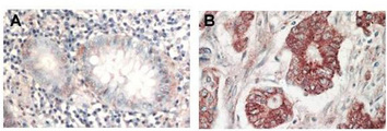
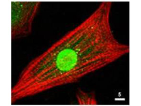
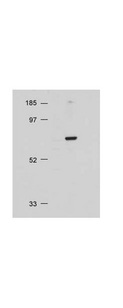
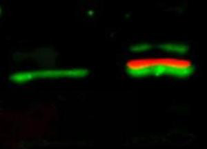
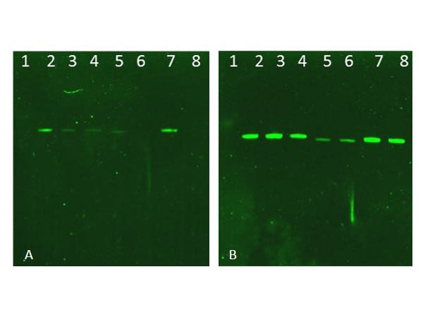
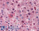
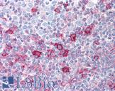
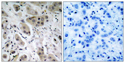
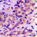
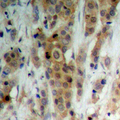
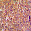
Polyclonal Rabbit anti‑Human AKT1 + AKT2 + AKT3 Antibody (aa460‑480, IHC, IF, WB) LS‑C744594
Polyclonal Rabbit anti‑Human AKT1 + AKT2 + AKT3 Antibody (aa460‑480, IHC, IF, WB) LS‑C744594
Antibody:
AKT1 + AKT2 + AKT3 Rabbit anti-Human Polyclonal (aa460-480) Antibody
Application:
IHC, IF, WB, Flo, ELISA
Reactivity:
Human, Mouse, Rat, Chicken
Format:
Unconjugated, Unmodified
Toll Free North America
 206-374-1102
For Research Use Only
Overview
Antibody:
AKT1 + AKT2 + AKT3 Rabbit anti-Human Polyclonal (aa460-480) Antibody
Application:
IHC, IF, WB, Flo, ELISA
Reactivity:
Human, Mouse, Rat, Chicken
Format:
Unconjugated, Unmodified
Specifications
Description
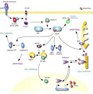
AKT1 + AKT2 + AKT3 antibody LS-C744594 is an unconjugated rabbit polyclonal antibody to AKT1 + AKT2 + AKT3 (aa460-480) from human. It is reactive with human, mouse, rat and other species. Validated for ELISA, Flow, IF, IHC and WB.
Host
Rabbit
Reactivity
Human, Mouse, Rat, Chicken
(tested or 100% immunogen sequence identity)
Clonality
Polyclonal
Conjugations
Unconjugated
Purification
Delipidated and defibrinated
Modifications
Unmodified
Immunogen
AKT Antibody was produced from whole rabbit serum prepared by repeated immunizations with a synthetic peptide R-P-H-F-P-Q-F-S-Y-S-A-S-G-T-A corresponding to the C-terminus (460-480) of human AKT proteins conjugated to KLH using maleimide. A residue of cysteine was added to the amino terminal end to facilitate coupling. A BLAST analysis was used to suggest reactivity with this protein from rat, mouse, and chicken based on 100% homology for the immunogen sequence.
Epitope
aa460-480
Specificity
Reacts with the AKT from human tissues. Based on sequence we expect this antibody to react as well with rat, mouse, and chicken AKT.
Applications
- IHC (1:500 - 1:2000)
- Immunofluorescence (1:100 - 1:1000)
- Western blot (1:500 - 1:2000)
- Flow Cytometry
- ELISA (1:2000 - 1:10000)

|
Performing IHC? See our complete line of Immunohistochemistry Reagents including antigen retrieval solutions, blocking agents
ABC Detection Kits and polymers, biotinylated secondary antibodies, substrates and more.
|
Usage
Applications should be user optimized.
Presentation
0.02 M Potassium Phosphate, pH 7.2, 0.15 M NaCl, 0.1% Sodium Azide
Storage
Store vial at -20°C or below prior to opening. Dilute 1:10 to minimize loss. Store the vial at -20°C or below after dilution. Avoid freeze-thaw cycles.
Restrictions
For research use only. Intended for use by laboratory professionals.
Publications (0)
Customer Reviews (0)
Featured Products
Species:
Human, Mouse, Rat, Chicken
Applications:
IHC, IHC - Paraffin, ICC, Immunofluorescence, Western blot, Flow Cytometry, ELISA
Species:
Human, Mouse, Rat
Applications:
IHC, IHC - Paraffin, Immunofluorescence, Western blot, ELISA
Species:
Human, Mouse, Rat
Applications:
IHC, Western blot, Peptide Enzyme-Linked Immunosorbent Assay
Species:
Human, Mouse, Rat, Bovine, Sheep
Applications:
IHC, IHC - Paraffin, ICC, Immunofluorescence, Western blot
Species:
Human, Mouse, Rat, Bovine, Chicken, Zebrafish
Applications:
IHC, IHC - Paraffin, ICC, Immunofluorescence, Western blot
Species:
Human, Mouse, Rat, Bovine, Dog, Chicken, Zebrafish
Applications:
IHC, IHC - Paraffin, Western blot
Request SDS/MSDS
To request an SDS/MSDS form for this product, please contact our Technical Support department at:
Technical.Support@LSBio.com
Requested From: United States
Date Requested: 4/27/2024
Date Requested: 4/27/2024


