Login
Registration enables users to use special features of this website, such as past
order histories, retained contact details for faster checkout, review submissions, and special promotions.
order histories, retained contact details for faster checkout, review submissions, and special promotions.
Forgot password?
Registration enables users to use special features of this website, such as past
order histories, retained contact details for faster checkout, review submissions, and special promotions.
order histories, retained contact details for faster checkout, review submissions, and special promotions.
Quick Order
Products
Antibodies
ELISA and Assay Kits
Research Areas
Infectious Disease
Resources
Purchasing
Reference Material
Contact Us
Locations
Orders Processing,
Shipping & Receiving,
Warehouse
2 Shaker Rd Suites
B001/B101
Shirley, MA 01464
Production Lab
Floor 6, Suite 620
20700 44th Avenue W
Lynnwood, WA 98036
Telephone Numbers
Tel: +1 (206) 374-1102
Fax: +1 (206) 577-4565
Contact Us
Additional Contact Details
Login
Registration enables users to use special features of this website, such as past
order histories, retained contact details for faster checkout, review submissions, and special promotions.
order histories, retained contact details for faster checkout, review submissions, and special promotions.
Forgot password?
Registration enables users to use special features of this website, such as past
order histories, retained contact details for faster checkout, review submissions, and special promotions.
order histories, retained contact details for faster checkout, review submissions, and special promotions.
Quick Order
| Catalog Number | Size | Price |
|---|---|---|
| LS-B16021-0.1 | 0.1 mg (1 mg/ml) | $485 |
![PD-L2 / PDCD1LG2 / CD273 Antibody - Immunofluorescence of PD-L2 in transfected HEK293 cells with PD-L2 antibody at 20 ug/mL. Green: PDL2 Antibody [10H6] Blue: DAPI staining](https://lsbio-7d62.kxcdn.com/image2/pathplus-pd-l2-pdcd1lg2-cd273-antibody-clone-10h6-ls-b16021/484793_3816980.gif)
![PD-L2 / PDCD1LG2 / CD273 Antibody - Immunofluorescence of PD-L2 in human colon carcinoma tissue with PD-L2 antibody at 20 ug/mL. Green: PDL2 Antibody [10H6] Blue: DAPI staining](https://lsbio-7d62.kxcdn.com/image2/pathplus-pd-l2-pdcd1lg2-cd273-antibody-clone-10h6-ls-b16021/484792_3816964.gif)
![PD-L2 / PDCD1LG2 / CD273 Antibody - Immunofluorescence of PD-L2 in human tonsil tissue with PD-L2 antibody at 20 ug/mL. Green: PDL2 Antibody [10H6] Blue: DAPI staining](https://lsbio-7d62.kxcdn.com/image2/pathplus-pd-l2-pdcd1lg2-cd273-antibody-clone-10h6-ls-b16021/484791_3816940.gif)
1 of 7
2 of 7
3 of 7
4 of 7
5 of 7
6 of 7
7 of 7
PathPlus™ Monoclonal Mouse anti‑Human PD‑L2 / PDCD1LG2 / CD273 Antibody (clone 10H6, IHC, IF, WB) LS‑B16021
PathPlus™ Monoclonal Mouse anti‑Human PD‑L2 / PDCD1LG2 / CD273 Antibody (clone 10H6, IHC, IF, WB) LS‑B16021

Note: This antibody replaces LS-C669074
Antibody:
PD-L2 / PDCD1LG2 / CD273 Mouse anti-Human Monoclonal (10H6) Antibody
Application:
IHC-P, ICC, IF, WB, Flo, ELISA
Reactivity:
Human
Format:
Unconjugated, Unmodified
Toll Free North America
 206-374-1102
For Research Use Only
Overview
Antibody:
PD-L2 / PDCD1LG2 / CD273 Mouse anti-Human Monoclonal (10H6) Antibody
Application:
IHC-P, ICC, IF, WB, Flo, ELISA
Reactivity:
Human
Format:
Unconjugated, Unmodified
Specifications
Description
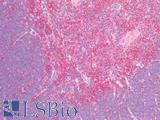
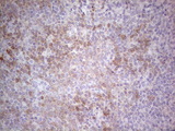
PDCD1LG (PDL2, CD273, programmed cell death 1 ligand 2) is a ligand for the programmed death-1 receptor (PD1), which is part of the inhibitory T-cell response (immune checkpoint) pathway and a molecule involved in peripheral tolerance and immune escape mechanisms during chronic viral infections and cancer. PDL2 functions in the induction and effector phases of T-cell immunity, dampening and regulating T-cell responsiveness, and inhibiting T cell proliferation, cytokine production, and cell adhesions. It is expressed in a variety of tissues and cancers, and along with PD1, a target of immunotherapy designed to block inhibitory receptors that are upregulated on tumor-associated T cells (immune checkpoint blockade).
References: Clin Dev Immunol 2012 ID656340; Int immunol 2010 22(*):651; Clin Can Res 2017 23(12):3158.
Target
Human PD-L2 / PDCD1LG2 / CD273
Synonyms
PDCD1LG2 | B7 dendritic cell molecule | B7-DC | B7DC | BA574F11.2 | CD273 | Btdc | CD273 antigen | PD-L2 | PDCD1 ligand 2 | PD-1-ligand 2 | Programmed death ligand 2 | Butyrophilin B7-DC | PD-1 ligand 2 | PDCD1L2 | PDL2
Host
Mouse
Reactivity
Human
(tested or 100% immunogen sequence identity)
Clonality
IgG1
Monoclonal
Clone
10H6
Conjugations
Unconjugated
Purification
Protein A purified
Modifications
Unmodified
Immunogen
The extracellular domain of human PD-L2.
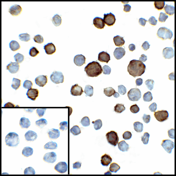
Applications
- IHC - Paraffin (10 µg/ml)
- ICC
- Immunofluorescence
- Western blot
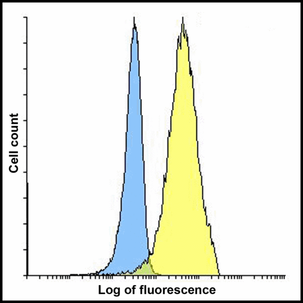
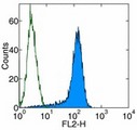
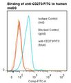
- Flow Cytometry
- ELISA

|
Performing IHC? See our complete line of Immunohistochemistry Reagents including antigen retrieval solutions, blocking agents
ABC Detection Kits and polymers, biotinylated secondary antibodies, substrates and more.
|
Usage
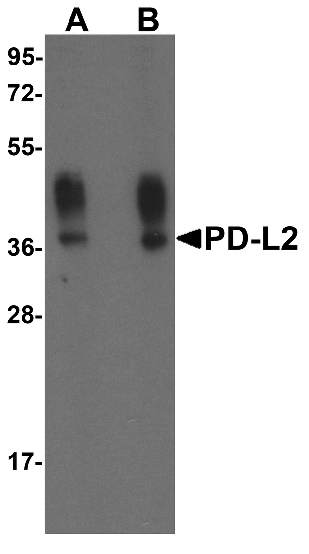
PD-L2 antibody can be used for detection of PD-L2 by Western blot at 0.5 - 1 ug/mL. Antibody can also be used for immunohistochemistry starting at 2 ug/mL. For immunofluorescence start at 20 ug/mL. Antibody validated: Western Blot in human samples; Immunohistochemistry in human samples; Immunocytochemistry in human samples; Immunofluorescence in human samples and Flow Cytometry in mouse samples. All other applications and species not yet tested. Western Blot: Predicted: 30 kDa Observed: 38 kDa
Presentation
PBS, 0.02% Sodium Azide, 50% Glycerol
Storage
Store at 4°C for up 3 months. Store at -20°C for up to 1 year. Avoid freeze/thaw cycles.
Restrictions
For research use only. Intended for use by laboratory professionals.
About PD-L2 / PDCD1LG2 / CD273
Validation
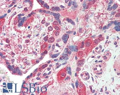
Human Placenta: Formalin-Fixed, Paraffin-Embedded (FFPE) at 10 µg/ml.
Human Placenta: Formalin-Fixed, Paraffin-Embedded (FFPE) at 10 µg/ml.
See More About...
LSBio Ratings
PathPlus™ PD-L2 / PDCD1LG2 / CD273 Antibody (clone 10H6) for IHC, ICC, IF/Immunofluorescence, WB/Western, Flow, ELISA LS-B16021 has an LSBio Rating of
Laboratory Validation Score (5)
Learn more about The LSBio Ratings Algorithm
Publications (0)
Customer Reviews (0)
Featured Products
Species:
Human
Applications:
IHC, IHC - Paraffin, Western blot
Species:
Mouse
Applications:
IHC, IHC - Frozen, Western blot, Immunoprecipitation, Flow Cytometry
Species:
Human, Mouse, Rat
Applications:
IHC, IHC - Paraffin, Western blot
Species:
Mouse
Applications:
IHC, IHC - Paraffin, IHC - Frozen, Immunoprecipitation, Flow Cytometry
Species:
Human
Applications:
Flow Cytometry, ELISA, Blocking
Request SDS/MSDS
To request an SDS/MSDS form for this product, please contact our Technical Support department at:
Technical.Support@LSBio.com
Requested From: United States
Date Requested: 4/27/2024
Date Requested: 4/27/2024


