Products
Research Areas
COVID-19
Resources
Login
Quick Order
Cart
Login
Registration enables users to use special features of this website, such as past
order histories, retained contact details for faster checkout, review submissions, and special promotions.


Fields marked with a * are required.

Login
Quick Order
Contact Us

Locations


Orders Processing,
Shipping & Receiving,
Warehouse

2 Shaker Rd Suites
B001/B101
Shirley, MA 01464


Production Lab

Floor 6, Suite 620
20700 44th Avenue W
Lynnwood, WA 98036

Telephone Numbers


Tel: +1 (206) 374-1102
Fax: +1 (206) 577-4565

Contact Us


Additional Contact Details

Login
Registration enables users to use special features of this website, such as past
order histories, retained contact details for faster checkout, review submissions, and special promotions.


Fields marked with a * are required.

Login
Quick Order
Catalog Number Size Price
LS-C796711-100 100 µg $539 
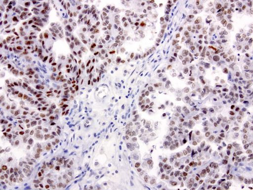
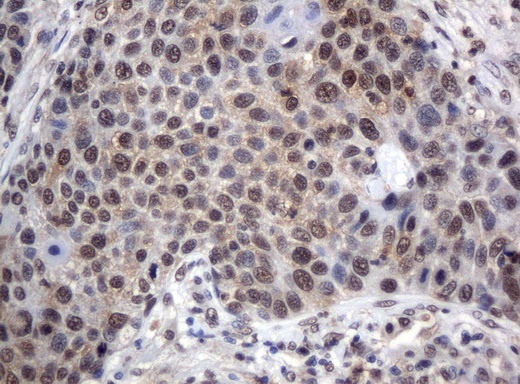
XRCC1 Antibody - Immunohistochemical staining of paraffin-embedded human ovarian carcinoma using anti-XRCC1 clone UMAB40 mouse monoclonal antibody  at 1:50 with Polink2 Broad HRP DAB detection kit; heat-induced epitope retrieval with GBI TEE pH9.0 HIER buffer using pressure chamber for 3 minutes at 110C. Nuclear staining is seen in tumor cells.
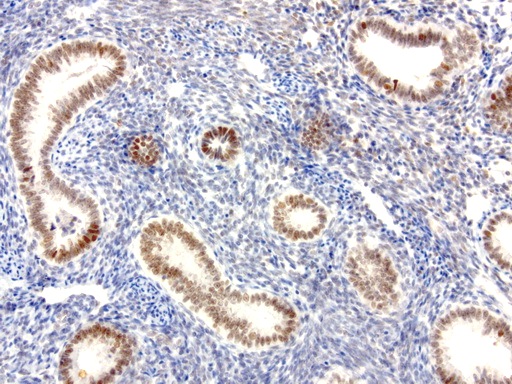
XRCC1 Antibody - Immunohistochemical staining of paraffin-embedded human endometrium using anti-XRCC1 clone UMAB40 mouse monoclonal antibody  at 1:50 with Polink2 Broad HRP DAB detection kit; heat-induced epitope retrieval with GBI TEE pH9.0 HIER buffer using pressure chamber for 3 minutes at 110C. Nuclear staining is seen in epithelial cells.
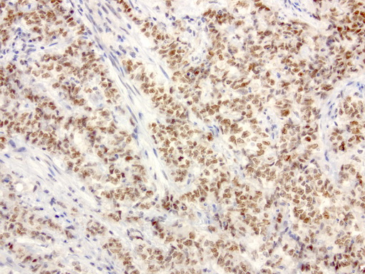
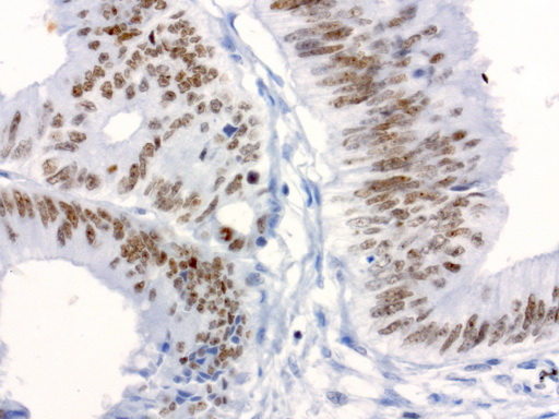
XRCC1 Antibody - Immunohistochemical staining of paraffin-embedded human endometrial carcinoma using anti-XRCC1 clone UMAB40 mouse monoclonal antibody  at 1:50 with Polink2 Broad HRP DAB detection kit; heat-induced epitope retrieval with GBI TEE pH9.0 HIER buffer using pressure chamber for 3 minutes at 110C. Nuclear staining is seen in tumor cells.
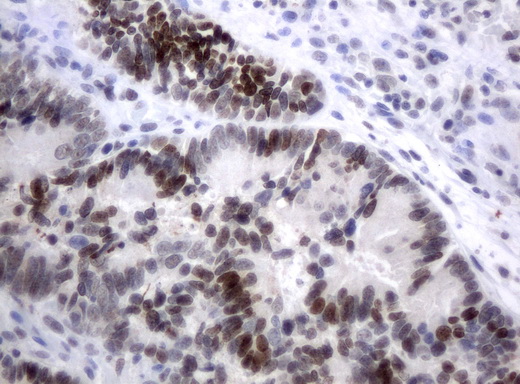
XRCC1 Antibody - Immunohistochemical staining of paraffin-embedded Adenocarcinoma of colon tissue using anti-XRCC1 mouse monoclonal antibody.  dilution 1:50; heat-induced epitope retrieval by 10mM citric buffer, pH6.0, 120C for 3min)
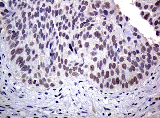
XRCC1 Antibody - Immunohistochemical staining of paraffin-embedded Carcinoma of bladder tissue using anti-XRCC1 mouse monoclonal antibody.  dilution 1:50; heat-induced epitope retrieval by 10mM citric buffer, pH6.0, 120C for 3min)
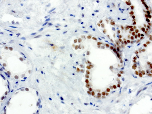
XRCC1 Antibody - Immunohistochemical staining of paraffin-embedded human prostate carcinoma using anti-XRCC1 clone UMAB40 mouse monoclonal antibody  at 1:50 with Polink2 Broad HRP DAB detection kit; heat-induced epitope retrieval with GBI TEE pH9.0 HIER buffer using pressure chamber for 3 minutes at 110C. Nuclear staining is seen in tumor cells.
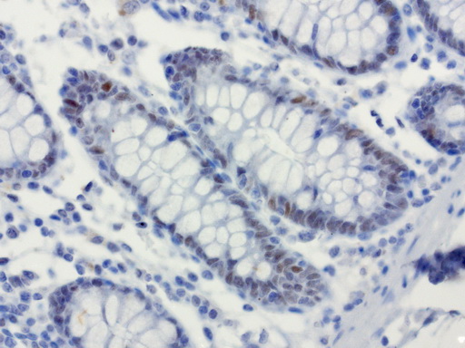
XRCC1 Antibody - Immunohistochemical staining of paraffin-embedded human colon using anti-XRCC1 clone UMAB40 mouse monoclonal antibody  at 1:50 with Polink2 Broad HRP DAB detection kit; heat-induced epitope retrieval with GBI Accel pH8.7 HIER buffer using pressure chamber for 3 minutes at 110C. Nuclear staining is seen in epithelial cells.
XRCC1 Antibody - Immunohistochemical staining of paraffin-embedded human colon carcinoma using anti-XRCC1 clone UMAB40 mouse monoclonal antibody  at 1:50 with Polink2 Broad HRP DAB detection kit; heat-induced epitope retrieval with GBI Accel pH8.7 HIER buffer using pressure chamber for 3 minutes at 110C. Nuclear staining is seen in tumor cells.
XRCC1 Antibody - Immunohistochemical staining of paraffin-embedded Carcinoma of lung tissue using anti-XRCC1 mouse monoclonal antibody.  dilution 1:50; heat-induced epitope retrieval by 10mM citric buffer, pH6.0, 120C for 3min)
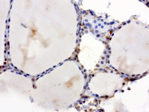
XRCC1 Antibody - Immunohistochemical staining of paraffin-embedded human thyroid using anti-XRCC1 clone UMAB40 mouse monoclonal antibody  at 1:50 with Polink2 Broad HRP DAB detection kit; heat-induced epitope retrieval with GBI TEE pH9.0 HIER buffer using pressure chamber for 3 minutes at 110C. Nuclear staining is seen in epithelial cells.
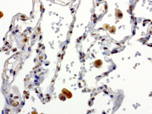
XRCC1 Antibody - Immunohistochemical staining of paraffin-embedded human lung using anti-XRCC1 clone UMAB40 mouse monoclonal antibody  at 1:50 with Polink2 Broad HRP DAB detection kit; heat-induced epitope retrieval with GBI TEE pH9.0 HIER buffer using pressure chamber for 3 minutes at 110C. Nuclear staining is seen in epithelial cells.
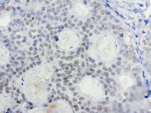
XRCC1 Antibody - Immunohistochemical staining of paraffin-embedded human breast carcinoma using anti-XRCC1 clone UMAB40 mouse monoclonal antibody  at 1:50 with Polink2 Broad HRP DAB detection kit; heat-induced epitope retrieval with GBI Accel pH8.7 HIER buffer using pressure chamber for 3 minutes at 110C. Weak nuclear staining is seen in tumor cells.
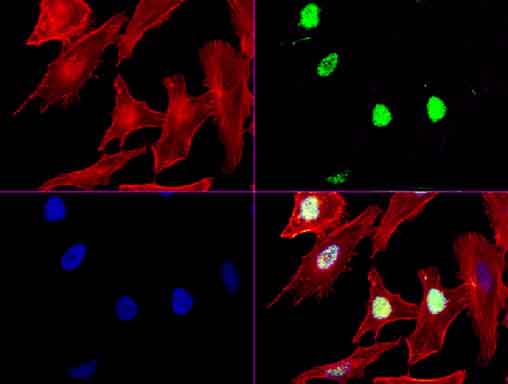
XRCC1 Antibody - Immunofluorescent staining of HeLa cells using anti-XRCC1 mouse monoclonal antibody  green). Actin filaments were labeled with TRITC-phalloidin. (red), and nuclear with DAPI. (blue). The three-color overlay image is located at the bottom-right corner.
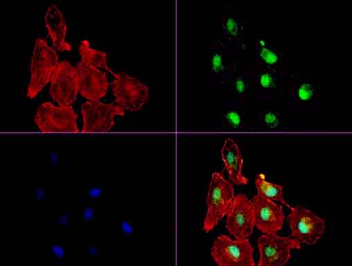
XRCC1 Antibody - Immunofluorescent staining of A549 cells using anti-XRCC1 mouse monoclonal antibody  green). Actin filaments were labeled with TRITC-phalloidin. (red), and nuclear with DAPI. (blue). The three-color overlay image is located at the bottom-right corner.
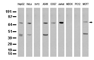
XRCC1 Antibody - Western blot analysis of extracts. (35ug) from 9 different cell lines by using anti-XRCC1 monoclonal antibody. (Clone UMAB40) at 1:500.
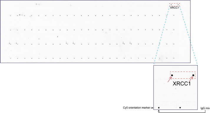
XRCC1 Antibody - OriGene overexpression protein microarray chip was immunostained with UltraMAB anti-XRCC1 mouse monoclonal antibody. The positive reactive proteins are highlighted with two red arrows in the enlarged subarray. All the positive controls spotted in this subarray are also labeled for clarification. These data show that UltraMAB anti-XRCC1. (Clone UMAB37) very specifically recognizes XRCC1 antigen on OriGene protein microarray chip.
1 of 16
2 of 16
3 of 16
4 of 16
5 of 16
6 of 16
7 of 16
8 of 16
9 of 16
10 of 16
11 of 16
12 of 16
13 of 16
14 of 16
15 of 16
16 of 16

Monoclonal Mouse anti‑Human XRCC1 Antibody (clone UMAB40, Carrier‑free, IHC, IF, WB) LS‑C796711

Monoclonal Mouse anti‑Human XRCC1 Antibody (clone UMAB40, Carrier‑free, IHC, IF, WB) LS‑C796711

Antibody:
XRCC1 Mouse anti-Human Monoclonal (Carrier-free) (UMAB40) Antibody
Application:
IHC, IF, WB, PMA
Reactivity:
Human, Monkey
Format:
Unconjugated, Carrier-free
Other formats:
Price
Catalog Number
$539
LS-C796711-100
Toll Free North America
206-374-1102
For Research Use Only

Overview

Antibody:
XRCC1 Mouse anti-Human Monoclonal (Carrier-free) (UMAB40) Antibody
Application:
IHC, IF, WB, PMA
Reactivity:
Human, Monkey
Format:
Unconjugated, Carrier-free
Other formats:

Specifications

Description
XRCC1 antibody LS-C796711 is an unconjugated mouse monoclonal antibody to XRCC1 from human. It is reactive with human and monkey. Validated for IF, IHC, PMA and WB.
Target
Human XRCC1
Synonyms
XRCC1 | RCC | DNA repair protein XRCC1
Host
Mouse
Reactivity
Human, Monkey (tested or 100% immunogen sequence identity)
Clonality
IgG1 Monoclonal
Clone
UMAB40
Conjugations
Unconjugated
Purification
Purified from ascites.
Modifications
Carrier-free. Also available Unmodified.
Immunogen
Full length human recombinant protein of human XRCC1 (NP_006288) produced in HEK293T cell.
Specificity
The specificity of this antibody has been validated by testing against a high-density Protein Microarray containing more than 17,000 recombinant proteins.
Applications
  • IHC (1:50)
  • Immunofluorescence (1:100)
  • Western blot
  • Protein Microarray
Performing IHC? See our complete line of Immunohistochemistry Reagents including antigen retrieval solutions, blocking agents ABC Detection Kits and polymers, biotinylated secondary antibodies, substrates and more.
Usage
Applications should be user optimized.
Presentation
PBS, pH 7.3, 8% Trehalose
Reconstitution
Reconstitute with PBS pH 7.3. To use this carrier-free antibody for conjugation experiment, we strongly recommend you to perform another round of desalting process.
Storage
Store at -20°C. Avoid freeze-thaw cycles.
Restrictions
For research use only. Intended for use by laboratory professionals.
Guarantee
This antibody carries the LSBio 100% Guarantee.
LSBio Guarantee
About XRCC1
P18887 NM_006297 NP_006288.2

Publications (0)

Customer Reviews (0)


Request SDS/MSDS

To request an SDS/MSDS form for this product, please contact our Technical Support department at:

Technical.Support@LSBio.com

Requested From: United States
Date Requested: 4/28/2024