Login
Registration enables users to use special features of this website, such as past
order histories, retained contact details for faster checkout, review submissions, and special promotions.
order histories, retained contact details for faster checkout, review submissions, and special promotions.
Forgot password?
Registration enables users to use special features of this website, such as past
order histories, retained contact details for faster checkout, review submissions, and special promotions.
order histories, retained contact details for faster checkout, review submissions, and special promotions.
Quick Order
Products
Antibodies
ELISA and Assay Kits
Research Areas
Infectious Disease
Resources
Purchasing
Reference Material
Contact Us
Locations
Orders Processing,
Shipping & Receiving,
Warehouse
2 Shaker Rd Suites
B001/B101
Shirley, MA 01464
Production Lab
Floor 6, Suite 620
20700 44th Avenue W
Lynnwood, WA 98036
Telephone Numbers
Tel: +1 (206) 374-1102
Fax: +1 (206) 577-4565
Contact Us
Additional Contact Details
Login
Registration enables users to use special features of this website, such as past
order histories, retained contact details for faster checkout, review submissions, and special promotions.
order histories, retained contact details for faster checkout, review submissions, and special promotions.
Forgot password?
Registration enables users to use special features of this website, such as past
order histories, retained contact details for faster checkout, review submissions, and special promotions.
order histories, retained contact details for faster checkout, review submissions, and special promotions.
Quick Order
| Catalog Number | Size | Price |
|---|---|---|
| LS-C756540-10 | 10 µg | $318 |
| LS-C756540-100 | 100 µg | $470 |
1 of 16
2 of 16
3 of 16
4 of 16
5 of 16
6 of 16
7 of 16
8 of 16
9 of 16
10 of 16
11 of 16
12 of 16
13 of 16
14 of 16
15 of 16
16 of 16
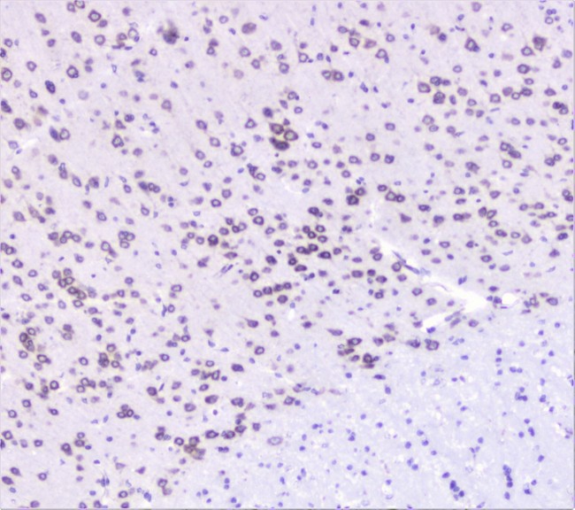
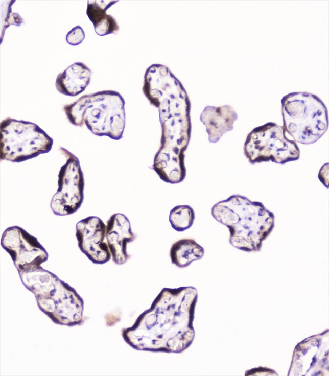
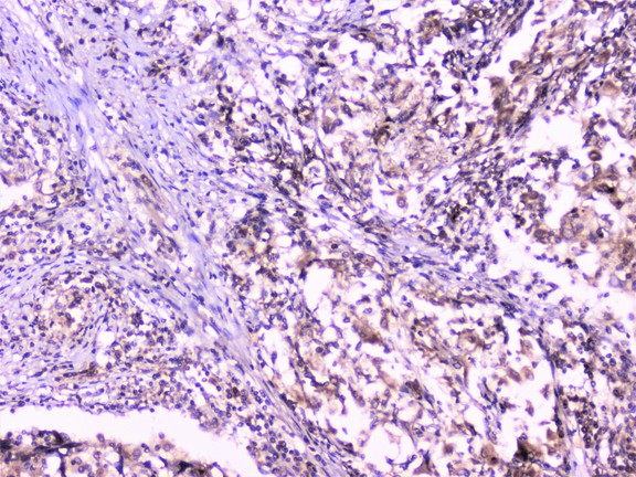
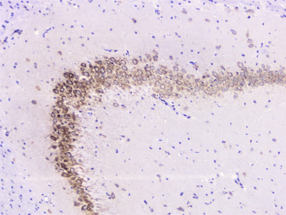
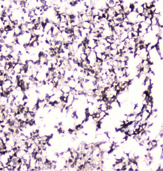
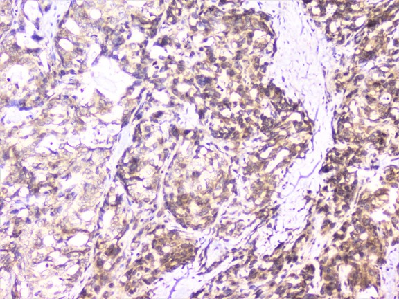
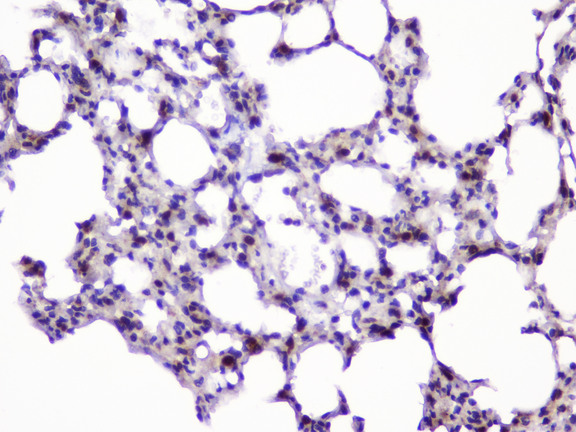
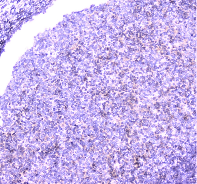
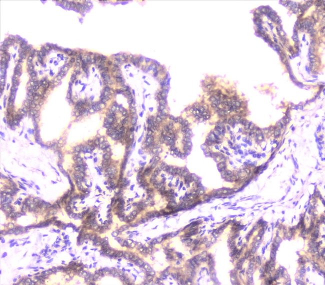
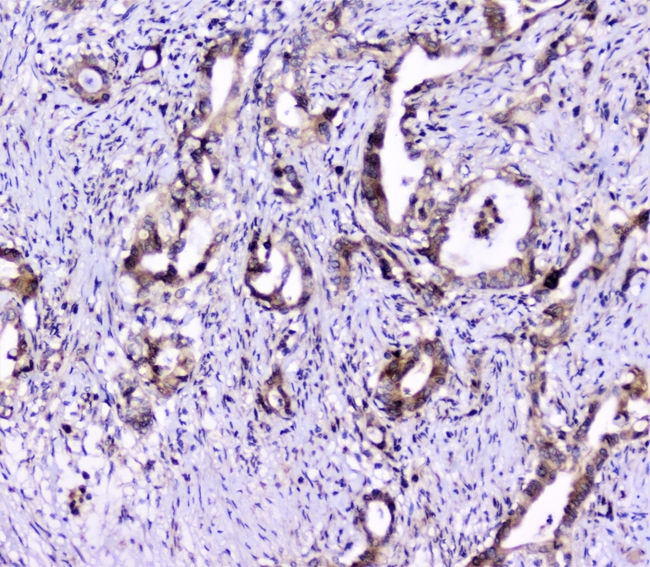
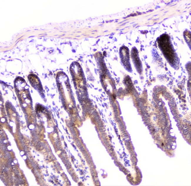
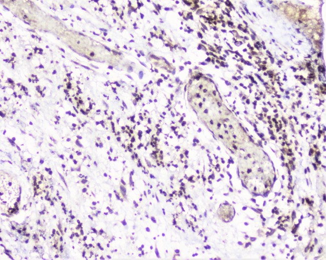
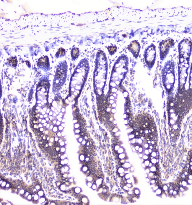
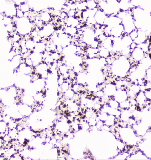
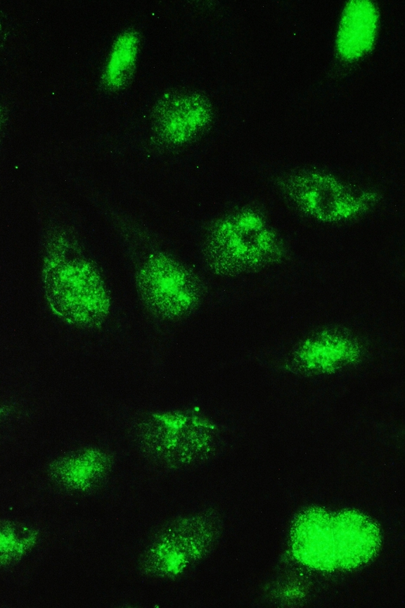
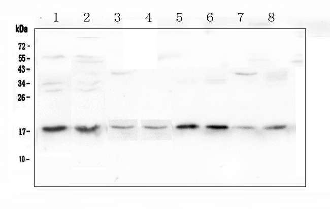
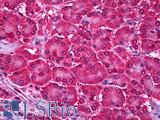
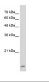
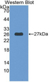
Polyclonal Rabbit anti‑Human UBE2I / UBC9 Antibody (IHC, WB) LS‑C756540
Polyclonal Rabbit anti‑Human UBE2I / UBC9 Antibody (IHC, WB) LS‑C756540
Antibody:
UBE2I / UBC9 Rabbit anti-Human Polyclonal Antibody
Application:
IHC-P, WB
Reactivity:
Human, Mouse, Rat
Format:
Unconjugated, Unmodified
Toll Free North America
 206-374-1102
For Research Use Only
Overview
Antibody:
UBE2I / UBC9 Rabbit anti-Human Polyclonal Antibody
Application:
IHC-P, WB
Reactivity:
Human, Mouse, Rat
Format:
Unconjugated, Unmodified
Specifications
Description
UBC9 antibody LS-C756540 is an unconjugated rabbit polyclonal antibody to UBC9 (UBE2I) from human. It is reactive with human, mouse and rat. Validated for IHC and WB.
Target
Human UBE2I / UBC9
Synonyms
UBE2I | C358B7.1 | SUMO-conjugating enzyme UBC9 | UBC9 | SUMO-protein ligase | UBCE9 | Ubiquitin carrier protein 9 | Ubiquitin carrier protein I | Ubiquitin-protein ligase I | p18 | SUMO-1-protein ligase | Ubiquitin conjugating enzyme 9 | Ubiquitin-protein ligase E2I
Host
Rabbit
Reactivity
Human, Mouse, Rat
(tested or 100% immunogen sequence identity)
Clonality
IgG
Polyclonal
Conjugations
Unconjugated
Purification
Immunogen affinity purified
Modifications
Unmodified
Immunogen
A synthetic peptide corresponding to a sequence of human UBE2I UBC9 (NIQDPAQAEAYTIYCQNRVEYEKRVRAQAKKFAPS).
Specificity
No cross reactivity with other proteins.
Applications
- IHC - Paraffin (0.5 - 1 µg/ml)
- Western blot (0.1 - 0.5 µg/ml)

|
Performing IHC? See our complete line of Immunohistochemistry Reagents including antigen retrieval solutions, blocking agents
ABC Detection Kits and polymers, biotinylated secondary antibodies, substrates and more.
|
Usage
Applications should be user optimized.
Presentation
Lyophilized from 0.2mg Na2HPO4, 0.9mg NaCl, 0.05mg sodium azide, 4mg Trehalose
Reconstitution
Add 0.2ml of distilled water will yield a concentration of 500µg/ml.
Storage
After reconstitution, may be stored at 4°C for 1 month. For long-term storage and to avoid freeze-thaw cycles, aliquot and store at -20°C.
Restrictions
For research use only. Intended for use by laboratory professionals.
About UBE2I / UBC9
Publications (0)
Customer Reviews (0)
Featured Products
Species:
Human, Monkey, Mouse, Rat, Bat, Bovine, Hamster, Horse, Rabbit, Chicken, Xenopus, Zebrafish
Applications:
IHC, IHC - Paraffin, Western blot, Peptide Enzyme-Linked Immunosorbent Assay

Reactivity:
Human
Range:
0.5-10 ng/ml
Request SDS/MSDS
To request an SDS/MSDS form for this product, please contact our Technical Support department at:
Technical.Support@LSBio.com
Requested From: United States
Date Requested: 4/28/2024
Date Requested: 4/28/2024


