Login
Registration enables users to use special features of this website, such as past
order histories, retained contact details for faster checkout, review submissions, and special promotions.
order histories, retained contact details for faster checkout, review submissions, and special promotions.
Forgot password?
Registration enables users to use special features of this website, such as past
order histories, retained contact details for faster checkout, review submissions, and special promotions.
order histories, retained contact details for faster checkout, review submissions, and special promotions.
Quick Order
Products
Antibodies
ELISA and Assay Kits
Research Areas
Infectious Disease
Resources
Purchasing
Reference Material
Contact Us
Locations
Orders Processing,
Shipping & Receiving,
Warehouse
2 Shaker Rd Suites
B001/B101
Shirley, MA 01464
Production Lab
Floor 6, Suite 620
20700 44th Avenue W
Lynnwood, WA 98036
Telephone Numbers
Tel: +1 (206) 374-1102
Fax: +1 (206) 577-4565
Contact Us
Additional Contact Details
Login
Registration enables users to use special features of this website, such as past
order histories, retained contact details for faster checkout, review submissions, and special promotions.
order histories, retained contact details for faster checkout, review submissions, and special promotions.
Forgot password?
Registration enables users to use special features of this website, such as past
order histories, retained contact details for faster checkout, review submissions, and special promotions.
order histories, retained contact details for faster checkout, review submissions, and special promotions.
Quick Order
| Catalog Number | Size | Price |
|---|---|---|
| LS-B10409-50 | 50 µl (1 mg/ml) | $460 |

1 of 3
2 of 3
3 of 3
IHC‑plus™ Monoclonal Mouse anti‑Human Rhodopsin / RHO Antibody (clone A531, IHC, IF, WB) LS‑B10409
IHC‑plus™ Monoclonal Mouse anti‑Human Rhodopsin / RHO Antibody (clone A531, IHC, IF, WB) LS‑B10409

Note: This antibody replaces LS-C204579
Antibody:
Rhodopsin / RHO Mouse anti-Human Monoclonal (A531) Antibody
Application:
IHC, IHC-P, IF, WB
Reactivity:
Human, Mouse, Rat, Bovine, Pig
Format:
Unconjugated, Unmodified
Toll Free North America
 206-374-1102
For Research Use Only
Overview
Antibody:
Rhodopsin / RHO Mouse anti-Human Monoclonal (A531) Antibody
Application:
IHC, IHC-P, IF, WB
Reactivity:
Human, Mouse, Rat, Bovine, Pig
Format:
Unconjugated, Unmodified
Specifications
Description
RHO antibody LS-B10409 is an unconjugated mouse monoclonal antibody to RHO (Rhodopsin) from human. It is reactive with human, mouse, rat and other species. Validated for IF, IHC and WB. Tested on 20 paraffin-embedded human tissues.
Target
Human Rhodopsin / RHO
Synonyms
RHO | CSNBAD1 | OPN2 | Rhodopsin | RP4 | Opsin 2, rod pigment | Opsin-2
Host
Mouse
Reactivity
Human, Mouse, Rat, Bovine, Pig
(tested or 100% immunogen sequence identity)
Clonality
IgG1,k
Monoclonal
Clone
A531
Conjugations
Unconjugated
Purification
Protein G purified
Modifications
Unmodified
Applications
- IHC
- IHC - Paraffin (10 µg/ml)
- Immunofluorescence (1:1000)
- Western blot (1:5000)

|
Performing IHC? See our complete line of Immunohistochemistry Reagents including antigen retrieval solutions, blocking agents
ABC Detection Kits and polymers, biotinylated secondary antibodies, substrates and more.
|
Usage
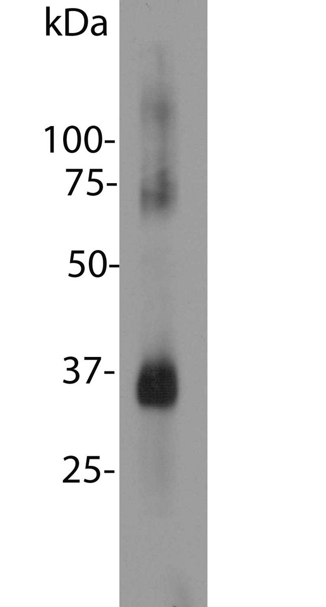
Try at dilutions of ~1:1000 for immunofluorescence. For western blots try at 1:5000. A suitable control tissue is retinal extracts. Rhodopsin run at 35 kDa on SDS-PAGE gels.
Presentation
PBS, 10 mM Sodium Azide
Storage
Store at 4°C or -20°C. Avoid freeze-thaw cycles.
Restrictions
For research use only. Intended for use by laboratory professionals.
About Rhodopsin / RHO
Validation
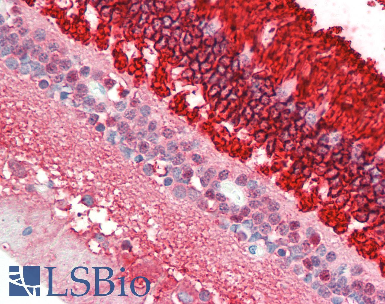
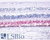
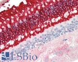
Anti-Rhodopsin / RHO antibody IHC staining of human retina. Immunohistochemistry of formalin-fixed, paraffin-embedded tissue after heat-induced antigen retrieval. Antibody concentration 10 ug/ml.
Anti-Rhodopsin / RHO antibody IHC staining of human retina. Immunohistochemistry of formalin-fixed, paraffin-embedded tissue after heat-induced antigen retrieval. Antibody concentration 10 ug/ml.
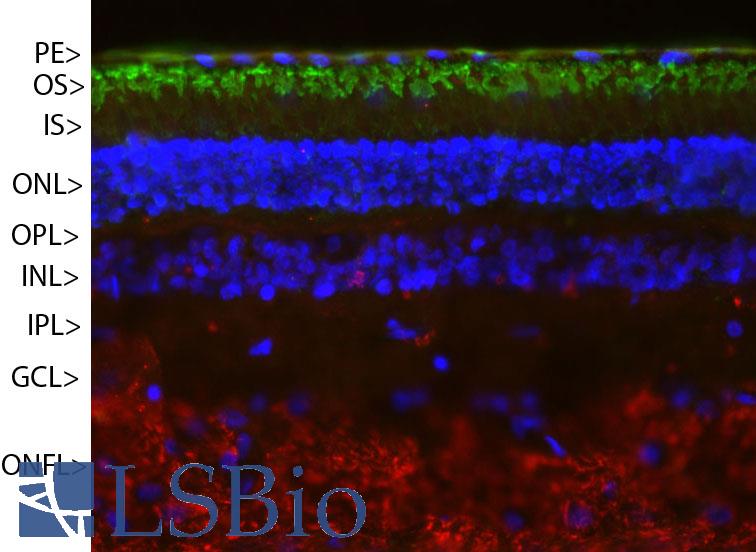
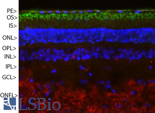
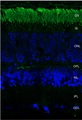
Pig retinal section stained with Rhodopsin / RHO antibody (green) and counterstained with rabbit polyclonal antibody to neurofilament RPCA-NF-M (red) and DNA (blue). Rhodopsin is most abundant in the outer segments of retina (OS), NF-M is abundant in the optic nerve fiber layer (ONFL), but seen in processes and neurons in other regions also. Other layers are pigmented epithelium (PE), outer and inner nuclear layers (ONL, INL), outer and inner plexiform layers (OPL, IPL) and ganglion cell layer (GCL).
Pig retinal section stained with Rhodopsin / RHO antibody (green) and counterstained with rabbit polyclonal antibody to neurofilament RPCA-NF-M (red) and DNA (blue). Rhodopsin is most abundant in the outer segments of retina (OS), NF-M is abundant in the optic nerve fiber layer (ONFL), but seen in processes and neurons in other regions also. Other layers are pigmented epithelium (PE), outer and inner nuclear layers (ONL, INL), outer and inner plexiform layers (OPL, IPL) and ganglion cell layer (GCL).
See More About...
LSBio Ratings
IHC-plus™ Rhodopsin / RHO Antibody (clone A531) for IHC, IF/Immunofluorescence, WB/Western LS-B10409 has an LSBio Rating of
Laboratory Validation Score (4)
Learn more about The LSBio Ratings Algorithm
Publications (0)
Customer Reviews (0)
Featured Products
Species:
Human, Monkey, Mouse, Rat, Pig, Rabbit
Applications:
IHC, IHC - Paraffin
Species:
Mammal, Human, Mouse, Rat, Bovine, Pig
Applications:
IHC, ICC, Immunofluorescence, Western blot, Immunoprecipitation, ELISA
Species:
Human, Mouse, Rat, Bovine, Pig
Applications:
IHC, IHC - Paraffin, ICC, Immunofluorescence, Western blot
Species:
Vertebrate
Applications:
IHC, ICC, Western blot, Immunoprecipitation, ELISA
Request SDS/MSDS
To request an SDS/MSDS form for this product, please contact our Technical Support department at:
Technical.Support@LSBio.com
Requested From: United States
Date Requested: 4/26/2024
Date Requested: 4/26/2024


