Products
Research Areas
COVID-19
Resources
Login
Quick Order
Cart Cart lightblue
Login
Registration enables users to use special features of this website, such as past
order histories, retained contact details for faster checkout, review submissions, and special promotions.


Fields marked with a * are required.

Login
Quick Order
Contact Us

Locations


Orders Processing,
Shipping & Receiving,
Warehouse

2 Shaker Rd Suites
B001/B101
Shirley, MA 01464


Production Lab

Floor 6, Suite 620
20700 44th Avenue W
Lynnwood, WA 98036

Telephone Numbers


Tel: +1 (800) 227-6666


Contact Us


Additional Contact Details

Login
Registration enables users to use special features of this website, such as past
order histories, retained contact details for faster checkout, review submissions, and special promotions.


Fields marked with a * are required.

Login
Quick Order

PLK1 / PLK-1

polo-like kinase 1

Serine/threonine-protein kinase that performs several important functions throughout M phase of the cell cycle, including the regulation of centrosome maturation and spindle assembly, the removal of cohesins from chromosome arms, the inactivation of anaphase-promoting complex/cyclosome (APC/C) inhibitors, and the regulation of mitotic exit and cytokinesis. Polo-like kinase proteins acts by binding and phosphorylating proteins are that already phosphorylated on a specific motif recognized by the POLO box domains. Phosphorylates BORA, BUB1B/BUBR1, CCNB1, CDC25C, CEP55, ECT2, ERCC6L, FBXO5/EMI1, FOXM1, KIF20A/MKLP2, CENPU, NEDD1, NINL, NPM1, NUDC, PKMYT1/MYT1, KIZ, PPP1R12A/MYPT1, PRC1, RACGAP1/CYK4, SGOL1, STAG2/SA2, TEX14, TOPORS, p73/TP73, TPT1 and WEE1. Plays a key role in centrosome functions and the assembly of bipolar spindles by phosphorylating KIZ, NEDD1 and NINL. NEDD1 phosphorylation promotes subsequent targeting of the gamma-tubulin ring complex (gTuRC) to the centrosome, an important step for spindle formation. Phosphorylation of NINL component of the centrosome leads to NINL dissociation from other centrosomal proteins. Involved in mitosis exit and cytokinesis by phosphorylating CEP55, ECT2, KIF20A/MKLP2, CENPU, PRC1 and RACGAP1. Recruited at the central spindle by phosphorylating and docking PRC1 and KIF20A/MKLP2; creates its own docking sites on PRC1 and KIF20A/MKLP2 by mediating phosphorylation of sites subsequently recognized by the POLO box domains. Phosphorylates RACGAP1, thereby creating a docking site for the Rho GTP exchange factor ECT2 that is essential for the cleavage furrow formation. Promotes the central spindle recruitment of ECT2. Plays a central role in G2/M transition of mitotic cell cycle by phosphorylating CCNB1, CDC25C, FOXM1, CENPU, PKMYT1/MYT1, PPP1R12A/MYPT1 and WEE1. Part of a regulatory circuit that promotes the activation of CDK1 by phosphorylating the positive regulator CDC25C and inhibiting the negative regulators WEE1 and PKMYT1/MYT1. Also acts by mediating phosphorylation of cyclin-B1 (CCNB1) on centrosomes in prophase. Phosphorylates FOXM1, a key mitotic transcription regulator, leading to enhance FOXM1 transcriptional activity. Involved in kinetochore functions and sister chromatid cohesion by phosphorylating BUB1B/BUBR1, FBXO5/EMI1 and STAG2/SA2. PLK1 is high on non-attached kinetochores suggesting a role of PLK1 in kinetochore attachment or in spindle assembly checkpoint (SAC) regulation. Required for kinetochore localization of BUB1B. Regulates the dissociation of cohesin from chromosomes by phosphorylating cohesin subunits such as STAG2/SA2. Phosphorylates SGOL1: required for spindle pole localization of isoform 3 of SGOL1 and plays a role in regulating its centriole cohesion function. Mediates phosphorylation of FBXO5/EMI1, a negative regulator of the APC/C complex during prophase, leading to FBXO5/EMI1 ubiquitination and degradation by the proteasome. Acts as a negative regulator of p53 family members: phosphorylates TOPORS, leading to inhibit the sumoylation of p53/TP53 and simultaneously enhance the ubiquitination and subsequent degradation of p53/TP53. Phosphorylates the transactivation domain of the transcription factor p73/TP73, leading to inhibit p73/TP73-mediated transcriptional activation and pro-apoptotic functions. Phosphorylates BORA, and thereby promotes the degradation of BORA. Contributes to the regulation of AURKA function. Also required for recovery after DNA damage checkpoint and entry into mitosis. Phosphorylates MISP, leading to stabilization of cortical and astral microtubule attachments required for proper spindle positioning.

Gene Name: polo-like kinase 1
Family/Subfamily: Protein Kinase , Polo
Synonyms: PLK1, Polo like kinase, Polo-like kinase (Drosophila), Polo-like kinase 1, STPK13, Polo (Drosophia)-like kinase, PLK, PLK-1
Target Sequences: NM_005030 NP_005021.2 P53350

Publications (5)

1
Arpc1b, a centrosomal protein, is both an activator and substrate of Aurora A. Molli PR, Li DQ, Bagheri-Yarmand R, Pakala SB, Katayama H, Sen S, Iyer J, Chernoff J, Tsai MY, Nair SS, Kumar R. The Journal of cell biology. 2010 190:101-14. [PubMed:20603326] [PMC:PMC2911675] Related Antibodies: LS-B1675.
2
Upregulation of FOXP1 is a new independent unfavorable prognosticator and a specific predictor of lymphatic dissemination in cutaneous melanoma patients. Donizy P, Pagacz K, Marczuk J, Fendler W, Maciejczyk A, Halon A, Matkowski R. OncoTargets and therapy. 2018 March;11:1413-1422. (Human) [Full Text Article] [PubMed:29559799] [PMC:PMC5857151] Related Antibodies: LS-C114491.
3
Polo-like kinase-1 immunoreactivity is associated with metastases in cutaneous melanoma. Maciej Kaczorowski, Tomasz Borowiec, Piotr Donizy, Konrad Pagacz, Wojciech Fendler, Artur Lipinski, Agnieszka Halon, Rafal Matkowski. Journal of cutaneous pathology. 2017 October;44:819-826. [Full Text Article] [PubMed:28626898] Related Antibodies: LS-C114491.
4
Disulfiram, a drug widely used to control alcoholism, suppresses the self-renewal of glioblastoma and over-rides resistance to temozolomide. Triscott J, Lee C, Hu K, Fotovati A, Berns R, Pambid M, Luk M, Kast RE, Kong E, Toyota E, Yip S, Toyota B, Dunn SE. Oncotarget. Oct;3:1112-23. [Full Text Article] [PubMed:23047041] [PMC:PMC3717961] Related Antibodies: LS-B4225.
5
Mechanism of the interaction of beta(2)-glycoprotein I with negatively charged phospholipid membranes. Hammel M, Schwarzenbacher R, Gries A, Kostner GM, Laggner P, Prassl R. Biochemistry. Nov;40:14173-81. [Full Text Article] [PubMed:11714270] Related Antibodies: LS-B50.
more

☰ Filters
Antibodies (89)
Proteins (8)
ELISA Kits (7)
Cell-Based (1)
Cell-Based Phosphorylation (1)
Custom (4)
Over-Expression Lysate (2)
Primary (89)
Recombinant (6)
Sandwich (1)
PLK1 / PLK-1 (104)
Human (92)
Mouse (34)
Rat (25)
Bat (3)
Bovine (3)
Chimpanzee (6)
Dog (9)
Gibbon (3)
Guinea pig (3)
Hamster (4)
Horse (3)
Macaque (1)
Monkey (8)
Pig (3)
Rabbit (3)
Xenopus (6)
Zebrafish (2)
IHC (39)
IHC-P (21)
WB (82)
Flo (11)
ELISA (41)
ICC (4)
IF (32)
IP (34)
Peptide-ELISA (5)
rabbit (47)
mouse (40)
goat (2)
Product Group
IHCPlus (6)
IgG (35)
IgG1 (10)
IgG1,k (14)
IgG1,k + IgG2b,k (6)
IgG2b (10)
monoclonal mc (40)
polyclonal pc (49)
1D1 (1)
1D4B (1)
2A3 (1)
2G12 (1)
3C11 (2)
3F8 (2)
OTI1A2 (2)
OTI1D4 (2)
OTI3F12 (2)
OTI5C1 (2)
OTI6C6 (2)
OTI8C12 (2)
OTI9E9 (2)
96-Well Microplate (2)
96-Well Strip Plate (5)
APC Conjugated (4)
Azide-free (1)
Biotin Conjugated (7)
Carrier-free (7)
FITC Conjugated (7)
HRP Conjugated (7)
PE Conjugated (4)
Unconjugated (60)
aa573-603 (7)
pThr210 (6)
C-Terminus (4)
N-Terminus (4)
150-230 aa (2)
80-160 aa (2)
Internal (2)
aa325-375 (2)
pSer137 (2)
Amino Acid: 331-508 (1)
aa1-50 (1)
aa100-200 (1)
aa176-225 (1)
aa287-298 (1)
aa300-603 (1)
aa402-603 (1)
aa86-430 (1)
p210 (1)
No (100)
Yes (4)
Adherent Cell Cultures (2)
Plasma (1)
Serum (1)
Tissue Homogenates (1)
6His, N-terminus (1)
GST (1)
GST, N-terminus (1)
His (1)
His,N-terminus (1)
Myc-DDK (Flag) (3)
Human (6)
Mouse (1)
293T Cells (1)
Baculovirus (2)
HEK 293 Cells (2)
Insect Cells (1)
Wheat Germ Extract (2)
Purified (4)
Low endotoxin level (2)
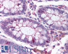
PLK1 / PLK-1 Antibody - Anti-PLK1 antibody IHC of human colon. Immunohistochemistry of formalin-fixed, paraffin-embedded tissue after heat-induced antigen retrieval. Antibody concentration 15 ug/ml.
Select
PLK1 / PLK-1 Mouse anti-Human Monoclonal (pThr210) (2A3) Antibody
Human
IHC, IHC-P, WB
Unconjugated
50 µg/$460
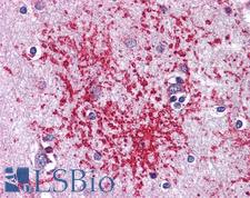
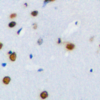
PLK1 / PLK-1 Antibody - Anti-PLK1 antibody IHC of human brain, cortex. Immunohistochemistry of formalin-fixed, paraffin-embedded tissue after heat-induced antigen retrieval. Antibody concentration 10 ug/ml.
Select
PLK1 / PLK-1 Rabbit anti-Human Polyclonal Antibody
Human
IHC, IHC-P, WB
Unconjugated
50 µl/$460
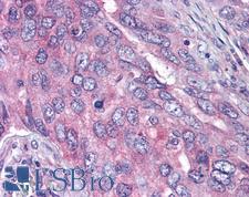
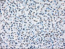
PLK1 / PLK-1 Antibody - Anti-PLK1 antibody IHC of human lung carcinoma. Immunohistochemistry of formalin-fixed, paraffin-embedded tissue after heat-induced antigen retrieval. Antibody concentration 10 ug/ml.
Select
PLK1 / PLK-1 Rabbit anti-Human Polyclonal (pThr210) Antibody
Human
ELISA, IHC, IHC-P, WB
Unconjugated
50 µg/$515
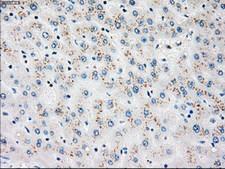
PLK1 / PLK-1 Antibody - IHC of paraffin-embedded liver tissue using anti-PLK1 mouse monoclonal antibody. (Dilution 1:50).
Select
PLK1 / PLK-1 Mouse anti-Human Monoclonal (OTI3F12) Antibody
Human
Flo, IHC, IHC-P, IP, WB
Unconjugated
100 µl/$434
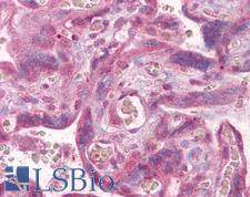
PLK1 / PLK-1 Antibody - Anti-PLK1 antibody IHC of human placenta. Immunohistochemistry of formalin-fixed, paraffin-embedded tissue after heat-induced antigen retrieval. Antibody dilution 1:100.
Select
PLK1 / PLK-1 Rabbit anti-Human Polyclonal (aa100-200) Antibody
Mouse, Dog, Bovine, Rat, Pig, Macaque, Human, Monkey
IHC, IHC-P, WB
Unconjugated
50 µl/$460
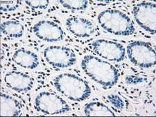
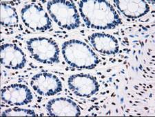
PLK1 / PLK-1 Antibody - IHC of paraffin-embedded colon tissue using anti-PLK1 mouse monoclonal antibody. (Dilution 1:50).
Select
PLK1 / PLK-1 Mouse anti-Human Monoclonal (OTI6C6) Antibody
Dog, Human
IHC, IHC-P, IP, WB
Unconjugated
100 µl/$434
PLK1 / PLK-1 Antibody - Immunohistochemical staining of paraffin-embedded breast tissue using anti-PLK1 mouse monoclonal antibody. (Dilution 1:50).
Select
PLK1 / PLK-1 Mouse anti-Human Monoclonal (OTI1A2) Antibody
IHC, IHC-P, WB
Unconjugated
100 µl/$434
PLK1 / PLK-1 Antibody - Immunohistochemical staining of paraffin-embedded breast tissue using anti-PLK1 mouse monoclonal antibody. (Dilution 1:50).
Select
PLK1 / PLK-1 Mouse anti-Human Monoclonal (Carrier-free) (OTI1A2) Antibody
Human
Flo, IHC, IP, WB
Unconjugated, Carrier-free
100 µg/$511
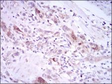
PLK1 / PLK-1 Antibody - IHC of paraffin-embedded stomach cancer tissues using PLK1 mouse monoclonal antibody with DAB staining.
Select
PLK1 / PLK-1 Mouse anti-Human Monoclonal (3C11) Antibody
Human
ELISA, Flo, IHC, WB
Unconjugated
100 µl/$459
PLK1 / PLK-1 Antibody - Detection of Human PLK1 by Immunocytochemistry. Sample: Formaldehyde-fixed asynchronous HeLa cells. Antibody: Affinity purified rabbit anti-PLK1 used at a dilution of 1:1000 (1 ug/ml). Detection: Green fluorescent goat anti-rabbit IgG highly cross-adsorbed Antibody FITC conjugated used at a dilution of 1:100.
Select
PLK1 / PLK-1 Rabbit anti-Human Polyclonal (aa325-375) Antibody
Human
ICC, IF, IP, WB
Unconjugated
10 µl/$295; 100 µl/$514
Select
PLK1 / PLK-1 Mouse anti-Human Monoclonal (HRP) Antibody
Human
ELISA, IF, IP, WB
HRP Conjugated
100 µl/$1,017
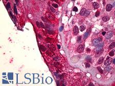
PLK1 / PLK-1 Antibody - Anti-PLK1 antibody IHC of human colon, epithelium. Immunohistochemistry of formalin-fixed, paraffin-embedded tissue after heat-induced antigen retrieval. Antibody concentration 5 ug/ml.
Select
PLK1 / PLK-1 Rabbit anti-Human Polyclonal Antibody
Human
IHC, IHC-P, WB
Unconjugated
50 µl/$460
Select
PLK1 / PLK-1 Rabbit anti-Human Polyclonal (aa573-603) (Biotin) Antibody
Human
ELISA, IHC, WB
Biotin Conjugated
200 µl/$952
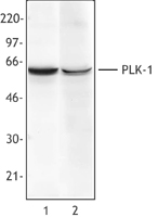
PLK1 / PLK-1 Antibody - Hela cell extract (Lane 1) or NIH3T3 cell extract (Lane 2) was resolved by electrophoresis, transferred to nitrocellulose and probed with monoclonal anti-Plk-1 (Clone 3F8) antibody. Proteins were visualized using a goat anti-mouse secondary conjugated to.
Select
PLK1 / PLK-1 Mouse anti-Human Monoclonal (3F8) Antibody
Mouse, Human
IP, WB
Unconjugated
25 µg/$279; 100 µg/$359
Select
PLK1 / PLK-1 Rabbit anti-Human Polyclonal (pSer137) Antibody
Mouse, Rat, Human
Peptide-ELISA, WB
Unconjugated
50 µl/$334; 100 µl/$397
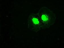
PLK1 / PLK-1 Antibody - Anti-PLK1 mouse monoclonal antibody  immunofluorescent staining of COS7 cells transiently transfected by pCMV6-ENTRY PLK1.
Select
PLK1 / PLK-1 Mouse anti-Human Monoclonal (OTI8C12) Antibody
Dog, Human, Monkey
Flo, IF, IP, WB
Unconjugated
100 µl/$434
PLK1 / PLK-1 Antibody - Anti-PLK1 mouse monoclonal antibody  immunofluorescent staining of COS7 cells transiently transfected by pCMV6-ENTRY PLK1.
Select
PLK1 / PLK-1 Mouse anti-Human Monoclonal (Carrier-free) (OTI8C12) Antibody
Dog, Human, Monkey
Flo, IF, IP, WB
Unconjugated, Carrier-free
100 µg/$511
PLK1 / PLK-1 Antibody - IHC of paraffin-embedded colon tissue using anti-PLK1 mouse monoclonal antibody. (Dilution 1:50).
Select
PLK1 / PLK-1 Mouse anti-Human Monoclonal (OTI5C1) Antibody
Human
Flo, IHC, IHC-P, IP, WB
Unconjugated
100 µl/$434
PLK1 / PLK-1 Antibody - IHC of paraffin-embedded colon tissue using anti-PLK1 mouse monoclonal antibody. (Dilution 1:50).
Select
PLK1 / PLK-1 Mouse anti-Human Monoclonal (Carrier-free) (OTI5C1) Antibody
Human
Flo, IHC, IP, WB
Unconjugated, Carrier-free
100 µg/$511
PLK1 / PLK-1 Antibody - Immunohistochemical analysis of PLK1 staining in human brain formalin fixed paraffin embedded tissue section. The section was pre-treated using heat mediated antigen retrieval with sodium citrate buffer (pH 6.0). The section was then incubated with the antibody at room temperature and detected using an HRP conjugated compact polymer system. DAB was used as the chromogen. The section was then counterstained with hematoxylin and mounted with DPX.
Select
PLK1 / PLK-1 Rabbit anti-Human Polyclonal (Internal) Antibody
Zebrafish, Pig, Mouse, Bovine, Human, Monkey
ICC, IF, IHC, IHC-P, WB
Unconjugated
30 µl/$255; 100 µl/$299; 200 µl/$351
PLK1 / PLK-1 Antibody - Immunofluorescence of monoclonal antibody to PLK1 on HeLa cell. [antibody concentration 10 ug/ml]
Select
PLK1 / PLK-1 Mouse anti-Human Monoclonal (2G12) Antibody
Human
ELISA, IF, IP, WB
Unconjugated
100 µg/$423
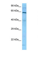
PLK1 / PLK-1 Antibody - PLK1 antibody Western blot of HeLa Cell lysate. Antibody concentration 1 ug/ml.  This image was taken for the unconjugated form of this product. Other forms have not been tested.
Select
PLK1 / PLK-1 Rabbit anti-Human Polyclonal (N-Terminus) Antibody
Human
WB
Unconjugated
100 µl/$424
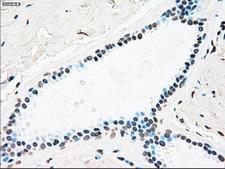
PLK1 / PLK-1 Antibody - IHC of paraffin-embedded pancreas tissue using anti-PLK1 mouse monoclonal antibody. (Dilution 1:50).
Select
PLK1 / PLK-1 Mouse anti-Human Monoclonal (OTI1D4) Antibody
Human
IHC, IHC-P, IP, WB
Unconjugated
100 µl/$434
PLK1 / PLK-1 Antibody - IHC of paraffin-embedded pancreas tissue using anti-PLK1 mouse monoclonal antibody. (Dilution 1:50).
Select
PLK1 / PLK-1 Mouse anti-Human Monoclonal (Carrier-free) (OTI1D4) Antibody
Human
Flo, IHC, IP, WB
Unconjugated, Carrier-free
100 µg/$511
Select
PLK1 / PLK-1 Rabbit anti-Human Polyclonal (150-230 aa) Antibody
Mouse, Rat, Human
ELISA, IF, IHC, WB
Unconjugated
50 µg/$295; 100 µg/$335; 200 µg/$394
Viewing 1-25 of 104 product results


If you do not find the reagent or information you require, please contact Customer.Support@LSBio.com to inquire about additional products in development.

PLEASE NOTE

For RESEARCH USE ONLY. Intended for use by laboratory professionals. Not intended for human diagnostic or therapeutic purposes.

The data on this page has been compiled from LifeSpan internal sources, the National Center for Biotechnology Information (NCBI), and The Universal Protein Resource (UniProt).