Login
Registration enables users to use special features of this website, such as past
order histories, retained contact details for faster checkout, review submissions, and special promotions.
order histories, retained contact details for faster checkout, review submissions, and special promotions.
Forgot password?
Registration enables users to use special features of this website, such as past
order histories, retained contact details for faster checkout, review submissions, and special promotions.
order histories, retained contact details for faster checkout, review submissions, and special promotions.
Quick Order
Products
Antibodies
ELISA and Assay Kits
Research Areas
Infectious Disease
Resources
Purchasing
Reference Material
Contact Us
Locations
Orders Processing,
Shipping & Receiving,
Warehouse
2 Shaker Rd Suites
B001/B101
Shirley, MA 01464
Production Lab
Floor 6, Suite 620
20700 44th Avenue W
Lynnwood, WA 98036
Telephone Numbers
Tel: +1 (206) 374-1102
Fax: +1 (206) 577-4565
Contact Us
Additional Contact Details
Login
Registration enables users to use special features of this website, such as past
order histories, retained contact details for faster checkout, review submissions, and special promotions.
order histories, retained contact details for faster checkout, review submissions, and special promotions.
Forgot password?
Registration enables users to use special features of this website, such as past
order histories, retained contact details for faster checkout, review submissions, and special promotions.
order histories, retained contact details for faster checkout, review submissions, and special promotions.
Quick Order
| Catalog Number | Size | Price |
|---|---|---|
| LS-B11179-50 | 50 µg (0.5 mg/ml) | $485 |
1 of 3
2 of 3
3 of 3
IHC‑plus™ Polyclonal Goat anti‑Human NOX1 Antibody (aa490‑502, IHC, WB) LS‑B11179
IHC‑plus™ Polyclonal Goat anti‑Human NOX1 Antibody (aa490‑502, IHC, WB) LS‑B11179

Note: This antibody replaces LS-C154813
Antibody:
NOX1 Goat anti-Human Polyclonal (aa490-502) Antibody
Application:
IHC, IHC-P, WB, Peptide-ELISA
Reactivity:
Human
Format:
Unconjugated, Unmodified
Toll Free North America
 206-374-1102
For Research Use Only
Overview
Antibody:
NOX1 Goat anti-Human Polyclonal (aa490-502) Antibody
Application:
IHC, IHC-P, WB, Peptide-ELISA
Reactivity:
Human
Format:
Unconjugated, Unmodified
Specifications
Description
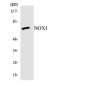
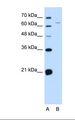
NOX1 antibody LS-B11179 is an unconjugated goat polyclonal antibody to human NOX1 (aa490-502). Validated for IHC, Peptide-ELISA and WB. Tested on 20 paraffin-embedded human tissues.
Target
Human NOX1
Synonyms
NOX1 | MOX1 | MOX-1 | NADPH oxidase 1 variant NOH-1L | NADPH oxidase homolog-1 | NOH-1 | NOH1 | Mitogenic oxidase 1 | NADPH oxidase 1 | gp91-2 | NOX-1
Host
Goat
Reactivity
Human
(tested or 100% immunogen sequence identity)
Clonality
Polyclonal
Conjugations
Unconjugated
Purification
Purified from goat serum by ammonium sulphate precipitation followed by antigen affinity chromatography using the immunizing peptide.
Modifications
Unmodified
Immunogen
Peptide with sequence C-DKATDIVTGLKQK, from the internal region of the protein sequence according to NP_008983.2NP_39249.1.
Epitope
aa490-502
Specificity
Human NOX1. This antibody is expected to recognize both reported isoforms (NP_008983.2; NP_39249.1).
Applications
- IHC
- IHC - Paraffin (5 µg/ml)
- Western blot (0.1 - 0.3 µg/ml)
- Peptide Enzyme-Linked Immunosorbent Assay (1:128000)

|
Performing IHC? See our complete line of Immunohistochemistry Reagents including antigen retrieval solutions, blocking agents
ABC Detection Kits and polymers, biotinylated secondary antibodies, substrates and more.
|
Usage
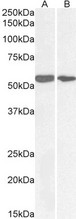
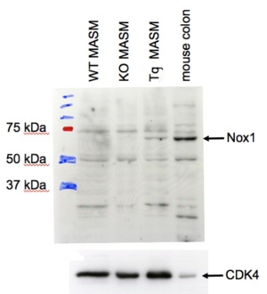
Peptide ELISA: antibody detection limit dilution 1:16000. Western blot: Approx 65kD band observed in Human and Mouse Colon lysates (calculated MW of 64.9kD according to NP_008983.2). The 65kD was also seen in cultured transgenic Mouse Aortic Smooth Muscle cells (MASM), but not to be seen at endogenous levels in MASM. Recommended concentration: 1-3 ug/ml.
Presentation
TBS, pH 7.3, 0.02% Sodium Azide, 0.5% BSA
Storage
Aliquot and store at -20°C. Avoid freeze-thaw cycles.
Restrictions
For research use only. Intended for use by laboratory professionals.
About NOX1
Validation
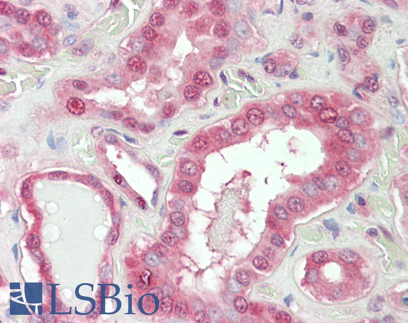
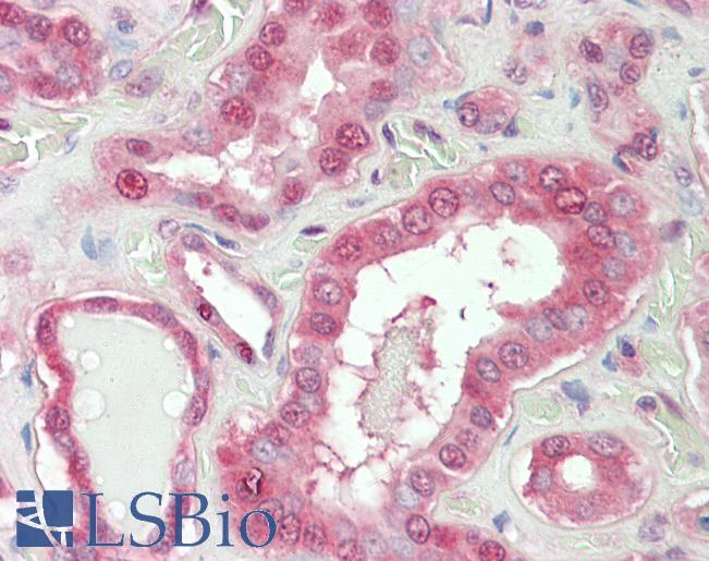
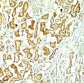
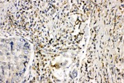
Anti-NOX1 antibody IHC staining of human kidney. Immunohistochemistry of formalin-fixed, paraffin-embedded tissue after heat-induced antigen retrieval. Antibody concentration 5 ug/ml.
Anti-NOX1 antibody IHC staining of human kidney. Immunohistochemistry of formalin-fixed, paraffin-embedded tissue after heat-induced antigen retrieval. Antibody concentration 5 ug/ml.
See More About...
LSBio Ratings
IHC-plus™ NOX1 Antibody (aa490-502) for IHC, WB/Western LS-B11179 has an LSBio Rating of
Laboratory Validation Score (4)
Learn more about The LSBio Ratings Algorithm
Publications (0)
Customer Reviews (0)
Featured Products
Species:
Human, Mouse
Applications:
Western blot, Peptide Enzyme-Linked Immunosorbent Assay
Species:
Human, Monkey
Applications:
IHC, IHC - Paraffin, Western blot
Request SDS/MSDS
To request an SDS/MSDS form for this product, please contact our Technical Support department at:
Technical.Support@LSBio.com
Requested From: United States
Date Requested: 4/26/2024
Date Requested: 4/26/2024


