Products
Research Areas
COVID-19
Resources
Login
Quick Order
Cart Cart lightblue
Login
Registration enables users to use special features of this website, such as past
order histories, retained contact details for faster checkout, review submissions, and special promotions.


Fields marked with a * are required.

Login
Quick Order
Contact Us

Locations


Orders Processing,
Shipping & Receiving,
Warehouse

2 Shaker Rd Suites
B001/B101
Shirley, MA 01464


Production Lab

Floor 6, Suite 620
20700 44th Avenue W
Lynnwood, WA 98036

Telephone Numbers


Tel: +1 (800) 227-6666


Contact Us


Additional Contact Details

Login
Registration enables users to use special features of this website, such as past
order histories, retained contact details for faster checkout, review submissions, and special promotions.


Fields marked with a * are required.

Login
Quick Order

CD3D

CD3d molecule, delta (CD3-TCR complex)

CD3D is part of the T-cell receptor/CD3 complex (TCR/CD3 complex) and is involved in T-cell development and signal transduction. The encoded membrane protein represents the delta subunit of the CD3 complex, and along with four other CD3 subunits, binds either TCR alpha/beta or TCR gamma/delta to form the TCR/CD3 complex on the surface of T-cells. Defects in this gene are a cause of severe combined immunodeficiency autosomal recessive T-cell-negative/B-cell-positive/NK-cell-positive (SCIDBNK). Two transcript variants encoding different isoforms have been found for this gene. Other variants may also exist, but the full-length natures of their transcripts has yet to be defined.

Gene Name: CD3d molecule, delta (CD3-TCR complex)
Synonyms: CD3D, CD3 delta, CD3d antigen, T-cell receptor T3 delta chain, T3D, CD3 antigen, delta subunit, CD3-DELTA, OKT3, delta chain
Target Sequences: NM_000732 NP_000723.1 P04234

☰ Filters
Antibodies (93)
Proteins (17)
ELISA Kits (16)
Custom (5)
Over-Expression Lysate (2)
Primary (93)
Recombinant (15)
Sandwich (10)
Sandwich CLIA (1)
CD3D (126)
Human (49)
Mouse (18)
Rat (21)
Bovine (9)
Chicken (2)
Cow (8)
Guinea pig (1)
Monkey (2)
Pig (9)
Porcine (1)
Rabbit (1)
Sheep (9)
IHC (7)
IHC-P (2)
WB (84)
Flo (5)
ELISA (12)
ICC (2)
IF (2)
IP (6)
Peptide-ELISA (3)
rabbit (72)
mouse (20)
goat (1)
IgG (29)
IgG1 (4)
IgG2a,k (4)
monoclonal mc (20)
polyclonal pc (73)
07 (1)
12 (1)
1F6A8 (1)
6 (1)
6B8D1G5 (1)
OKT3 (4)
12 x 8-Well Microstrips (1)
96-Well Microplate (1)
96-Well Strip Plate (14)
APC (7)
APC Conjugated (9)
Azide-free (1)
Biotin Conjugated (8)
Cy3 Conjugated (7)
Cy7 Conjugated (7)
FITC Conjugated (9)
HRP Conjugated (8)
PE Conjugated (9)
Preservative Free (7)
Unconjugated (36)
aa22-104 (24)
aa18-113 (8)
aa19-102 (8)
aa19-99 (8)
aa22-103 (8)
Internal (3)
Internal Region (1)
aa121-171 (1)
aa22-105 (1)
aa7-56 (1)
No (126)
Cell Lysates (1)
Plasma (4)
Serum (4)
Tissue Homogenates (2)
6His, C-terminus (1)
Fc, C-terminus (2)
Fc-DDK (Flag) (1)
GST (1)
GST, N-terminus (1)
His (5)
His,C-terminus (2)
His-GST (1)
His-S (1)
Myc-DDK (Flag) (2)
Bovine (1)
Crab-eating macaque (2)
Cynomolgus monkey (1)
Human (7)
Mouse (2)
Pig (1)
Rat (1)
Sheep (1)
293T Cells (1)
E. coli (6)
HEK 293 Cells (2)
Human Cells (3)
Mammalian Cells (3)
Wheat Germ Extract (2)
Purified (5)
Low endotoxin level (7)
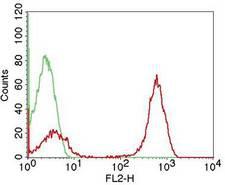
CD3D Antibody - Fig-1: Cell surface flow analysis of CD3 in PBMC using 0.1 µg/10^6 cells. Green represents isotype control; red represents anti-hCD3 antibody conjugated with PE.
Select
CD3D Mouse anti-Human Monoclonal (PE) (OKT3) Antibody
Human
Flo
PE Conjugated
25 Tests/$283; 100 Tests/$347
Select
CD3D Mouse anti-Human Monoclonal (1F6A8) Antibody
Human
ELISA, Flo, WB
Unconjugated
100 µl/$459
Select
CD3D Mouse anti-Human Monoclonal (FITC) (OKT3) Antibody
Human
Flo
FITC Conjugated
25 µg/$275; 100 µg/$323
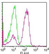
CD3D Antibody - Fig-1: Cell surface flow analysis of hCD3 in PBMC using 0.5 µg/10^6 cells. Green represents isotype control; red represents APC conjugated anti-hCD3 antibody (AC).
Select
CD3D Mouse anti-Human Monoclonal (APC) (OKT3) Antibody
Human
Flo
APC Conjugated
25 Tests/$274; 100 Tests/$347
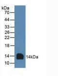
CD3D Antibody - Western blot of recombinant CD3D.  This image was taken for the unconjugated form of this product. Other forms have not been tested.
Select
CD3D Rabbit anti-Human Polyclonal (aa18-113) (FITC) Antibody
Human
WB
FITC Conjugated
100 µl/$341; 200 µl/$397; 500 µl/$583; 1 ml/$715; 2 ml/$953; 5 ml/$1,628
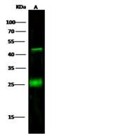
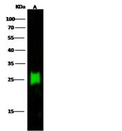
CD3D Antibody - Anti-CD3D rabbit monoclonal antibody at 1:500 dilution. Lane A: Jurkat Whole Cell Lysate. Lysates/proteins at 30 ug per lane. Secondary: Goat Anti-Rabbit IgG H&L (Dylight800) at 1/10000 dilution. Developed using the Odyssey technique. Performed under reducing conditions. Predicted band size: 19 kDa. Observed band size: 25 kDa. (We are unsure as to the identity of these extra bands.)
Select
CD3D Rabbit anti-Human Monoclonal (Preservative Free) (6) Antibody
Human
ELISA, IP, WB
Unconjugated, Preservative Free
100 µl/$412
CD3D Antibody
Select
CD3D Rabbit anti-Sheep Polyclonal (aa19-99) (Biotin) Antibody
Sheep
WB
Biotin Conjugated
100 µl/$343; 200 µl/$399; 500 µl/$587; 1 ml/$725; 2 ml/$963; 5 ml/$1,648
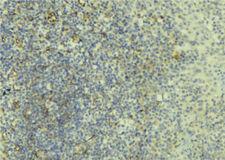
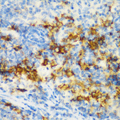
CD3D Antibody - 1:100 staining human lymph tissue by IHC-P. The sample was formaldehyde fixed and a heat mediated antigen retrieval step in citrate buffer was performed. The sample was then blocked and incubated with the antibody for 1.5 hours at 22°C. An HRP conjugated goat anti-rabbit antibody was used as the secondary.
Select
CD3D Rabbit anti-Human Polyclonal Antibody
Mouse, Rat, Human
IF, IHC, Peptide-ELISA, WB
Unconjugated
100 µl/$379; 200 µl/$421
CD3D ELISA Kit
Select
Sandwich
96-Well Microplate
Human
0.16 - 10 ng/ml
Colorimetric - 450nm (TMB)
Plasma, Serum
1 Plate/$586
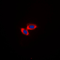
CD3D Antibody - Immunofluorescent analysis of CD3d staining in Jurkat cells. Formalin-fixed cells were permeabilized with 0.1% Triton X-100 in TBS for 5-10 minutes and blocked with 3% BSA-PBS for 30 minutes at room temperature. Cells were probed with the primary antibody in 3% BSA-PBS and incubated overnight at 4 C in a humidified chamber. Cells were washed with PBST and incubated with a DyLight 594-conjugated secondary antibody (red) in PBS at room temperature in the dark. DAPI was used to stain the cell nuclei (blue).
Select
CD3D Rabbit anti-Human Polyclonal (Internal) Antibody
Human, Monkey
ICC, IF, WB
Unconjugated
100 µl/$299
Select
CD3D Rabbit anti-Human Polyclonal Antibody
Human
ELISA, IHC, WB
Unconjugated
100 µg/$341
CD3D Antibody - Western blot of CD3-delta antibody
Select
CD3D Rabbit anti-Human Polyclonal (Internal) Antibody
Human
ELISA, ICC, WB
Unconjugated
50 µg/$295; 100 µg/$335; 200 µg/$394
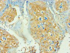
CD3D Antibody - Immunohistochemistry of paraffin-embedded human tonsil using antibody at 1:100 dilution.
Select
CD3D Rabbit anti-Human Polyclonal (aa22-105) Antibody
Human
ELISA, IHC, IHC-P
Unconjugated
50 µl/$294; 100 µl/$360
CD3D Antibody - Anti-CD3D mouse monoclonal antibody at 1:500 dilution. Lane A: Jurkat Whole Cell Lysate. Lysates/proteins at 30 ug per lane. Secondary: Goat Anti-Mouse IgG H&L (Dylight800) at 1/15000 dilution. Developed using the Odyssey technique. Performed under reducing conditions. Predicted band size: 19 kDa. Observed band size: 24 kDa.
Select
CD3D Mouse anti-Human Monoclonal (Preservative Free) (6B8D1G5) Antibody
Human
ELISA, IP, WB
Unconjugated, Preservative Free
20 µl/$256; 100 µl/$412
Select
CD3D Mouse anti-Human Monoclonal Antibody
Human
IHC, IHC-P
Unconjugated
100 µl/$412
Select
CD3D Rabbit anti-Human Polyclonal Antibody
Rat, Human
IHC, WB
Unconjugated
60 µl/$331; 120 µl/$403; 200 µl/$529
CD3D Antibody - Western blot of CD3D pAb in extracts from Jurkat and HuT-78 cells.
Select
CD3D Rabbit anti-Human Polyclonal Antibody
Mouse, Rat, Human
IHC, WB
Unconjugated
50 µl/$356; 100 µl/$461
Select
CD3D Mouse anti-Human Monoclonal Antibody
Human
ELISA, WB
Unconjugated
100 µg/$351
CD3D Antibody - Immunohistochemistry of paraffin-embedded rat spleen tissue.
Select
CD3D Rabbit anti-Human Polyclonal Antibody
Rat, Human
IHC, WB
Unconjugated
20 µl/$275; 100 µl/$389
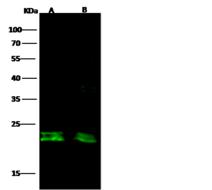
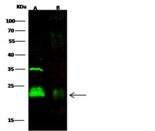
CD3D Antibody - Anti-CD3D rabbit polyclonal antibody at 1:500 dilution. Lane A: Jurkat Whole Cell Lysate. Lane B: Hu-T78 Whole Cell Lysate. Lysates/proteins at 20 ug per lane. Secondary: Goat Anti-Rabbit IgG H&L (Dylight800) at 1/10000 dilution. Developed using the Odyssey technique. Performed under reducing conditions. Predicted band size: 19 kDa. Observed band size: 21 kDa.
Select
CD3D Rabbit anti-Human Polyclonal (Preservative Free) Antibody
Human
ELISA, IP, WB
Unconjugated, Preservative Free
100 µl/$412
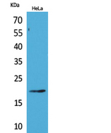
CD3D Antibody - Western Blot analysis of extracts from HeLa cells using CD3D Antibody.
Select
CD3D Rabbit anti-Human Polyclonal Antibody
Human
ELISA, WB
Unconjugated
50 µl/$334; 100 µl/$397
CD3D Antibody - Anti-CD3D mouse monoclonal antibody at 1:500 dilution. Lane A: Jurkat Whole Cell Lysate. Lane B: Molt-4 Whole Cell Lysate. Lysates/proteins at 30 ug per lane. Secondary: Goat Anti-Mouse IgG H&L (Dylight800) at 1/15000 dilution. Developed using the Odyssey technique. Performed under reducing conditions. Predicted band size: 19 kDa. Observed band size: 24 kDa.
Select
CD3D Mouse anti-Human Monoclonal (Preservative Free) (12) Antibody
Human
IP, WB
Unconjugated, Preservative Free
100 µl/$412
Select
CD3D Rabbit anti-Human Polyclonal Antibody
Human
IP, WB
Unconjugated
100 µl/$412
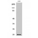
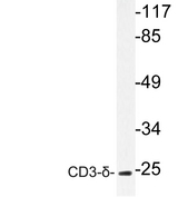
CD3D Antibody - Western blot analysis of lysate from A549 cells, using CD3-δ antibody.
Select
CD3D Rabbit anti-Human Polyclonal (aa7-56) Antibody
Human
Peptide-ELISA, WB
Unconjugated
50 µl/$334; 100 µl/$397
CD3D Antibody - Detection of human CD3D by western blot. Samples: Whole cell lysate (50 µg) from HeLa, HEK293T, and Jurkat cells prepared using NETN lysis buffer. Antibody: Affinity purified rabbit anti-CD3D antibody used for WB at 0.1 µg/ml. Detection: Chemiluminescence with an exposure time of 30 seconds.
Select
CD3D Rabbit anti-Human Polyclonal (aa121-171) Antibody
Human
IP, WB
Unconjugated
10 µl/$295; 100 µl/$514
Viewing 1-25 of 126 product results


If you do not find the reagent or information you require, please contact Customer.Support@LSBio.com to inquire about additional products in development.

PLEASE NOTE

For RESEARCH USE ONLY. Intended for use by laboratory professionals. Not intended for human diagnostic or therapeutic purposes.

The data on this page has been compiled from LifeSpan internal sources, the National Center for Biotechnology Information (NCBI), and The Universal Protein Resource (UniProt).