Login
Registration enables users to use special features of this website, such as past
order histories, retained contact details for faster checkout, review submissions, and special promotions.
order histories, retained contact details for faster checkout, review submissions, and special promotions.
Forgot password?
Registration enables users to use special features of this website, such as past
order histories, retained contact details for faster checkout, review submissions, and special promotions.
order histories, retained contact details for faster checkout, review submissions, and special promotions.
Quick Order
Products
Antibodies
ELISA and Assay Kits
Research Areas
Infectious Disease
Resources
Purchasing
Reference Material
Contact Us
Locations
Orders Processing,
Shipping & Receiving,
Warehouse
2 Shaker Rd Suites
B001/B101
Shirley, MA 01464
Production Lab
Floor 6, Suite 620
20700 44th Avenue W
Lynnwood, WA 98036
Telephone Numbers
Tel: +1 (206) 374-1102
Fax: +1 (206) 577-4565
Contact Us
Additional Contact Details
Login
Registration enables users to use special features of this website, such as past
order histories, retained contact details for faster checkout, review submissions, and special promotions.
order histories, retained contact details for faster checkout, review submissions, and special promotions.
Forgot password?
Registration enables users to use special features of this website, such as past
order histories, retained contact details for faster checkout, review submissions, and special promotions.
order histories, retained contact details for faster checkout, review submissions, and special promotions.
Quick Order
| Catalog Number | Size | Price |
|---|---|---|
| LS-C392589-200 | 200 µl | $393 |
1 of 3
2 of 3
3 of 3
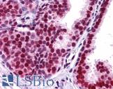
Monoclonal Mouse anti‑Human APEX1 / APE1 Antibody (clone 1518CT337.123.86.269.232, IHC, IF, WB) LS‑C392589
Monoclonal Mouse anti‑Human APEX1 / APE1 Antibody (clone 1518CT337.123.86.269.232, IHC, IF, WB) LS‑C392589

Antibody:
APEX1 / APE1 Mouse anti-Human Monoclonal (1518CT337.123.86.269.232) Antibody
Application:
IHC, IHC-P, IF, WB, Peptide-ELISA
Reactivity:
Human
Format:
Unconjugated, Unmodified
Toll Free North America
 206-374-1102
For Research Use Only
Overview
Antibody:
APEX1 / APE1 Mouse anti-Human Monoclonal (1518CT337.123.86.269.232) Antibody
Application:
IHC, IHC-P, IF, WB, Peptide-ELISA
Reactivity:
Human
Format:
Unconjugated, Unmodified
Specifications
Description
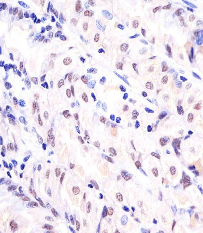
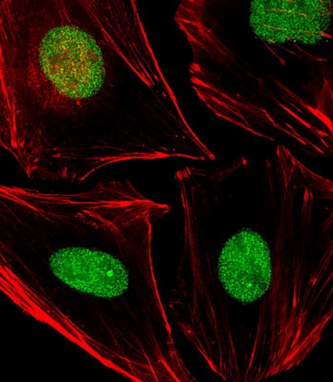
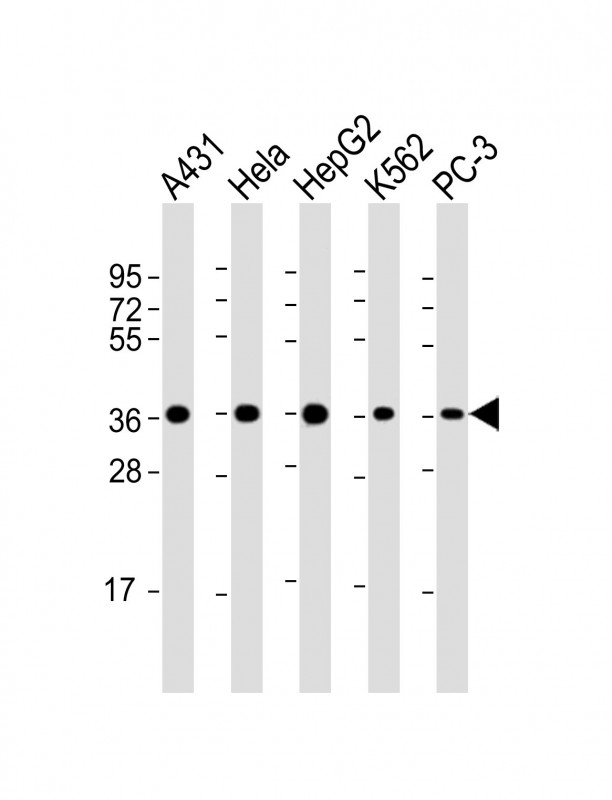
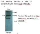
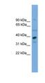
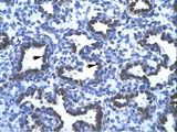
APE1 antibody LS-C392589 is an unconjugated mouse monoclonal antibody to human APE1 (APEX1). Validated for IF, IHC, Peptide-ELISA and WB. Cited in 1 publication.
Target
Human APEX1 / APE1
Synonyms
APEX1 | AP endonuclease class I | AP lyase | APE | APE-1 | APEN | APE1 | APEX nuclease | Redox factor-1 | AP endonuclease 1 | APEX | APX | Protein REF-1 | REF-1 | REF1
Host
Mouse
Reactivity
Human
(tested or 100% immunogen sequence identity)
Clonality
IgG1,k
Monoclonal
Clone
1518CT337.123.86.269.232
Conjugations
Unconjugated
Purification
Protein G purified
Modifications
Unmodified
Applications
- IHC
- IHC - Paraffin (1:25)
- Immunofluorescence (1:25)
- Western blot (1:2000)
- Peptide Enzyme-Linked Immunosorbent Assay

|
Performing IHC? See our complete line of Immunohistochemistry Reagents including antigen retrieval solutions, blocking agents
ABC Detection Kits and polymers, biotinylated secondary antibodies, substrates and more.
|
Presentation
PBS, 0.09% Sodium Azide
Storage
Maintain refrigerated at 2°C to 8°C for up to 6 months. For long term storage store at -20°C.
Restrictions
For research use only. Intended for use by laboratory professionals.
About APEX1 / APE1
LSBio Ratings
APEX1 / APE1 Antibody (clone 1518CT337.123.86.269.232) for IHC, IF/Immunofluorescence, WB/Western LS-C392589 has an LSBio Rating of
Publications (4)
Learn more about The LSBio Ratings Algorithm
Publications (1)
APEX1 Expression as a Potential Diagnostic Biomarker of Clear Cell Renal Cell Carcinoma and Hepatobiliary Carcinomas. Kim JM, Yeo MK, Lim JS, Song IS, Chun K, Kim KH. Journal of Clinical Medicine. 2019 August;8:E1151.
Customer Reviews (0)
Featured Products
Species:
Human, Mouse, Rat
Applications:
IHC, IHC - Frozen, ICC, Western blot, Immunoprecipitation
Species:
Human
Applications:
IHC, IHC - Paraffin, Western blot, Peptide Enzyme-Linked Immunosorbent Assay
Species:
Human
Applications:
IHC, IHC - Paraffin, Western blot
Request SDS/MSDS
To request an SDS/MSDS form for this product, please contact our Technical Support department at:
Technical.Support@LSBio.com
Requested From: United States
Date Requested: 4/25/2024
Date Requested: 4/25/2024


