order histories, retained contact details for faster checkout, review submissions, and special promotions.
Forgot password?
order histories, retained contact details for faster checkout, review submissions, and special promotions.
Locations
Orders Processing,
Shipping & Receiving,
Warehouse
2 Shaker Rd Suites
B001/B101
Shirley, MA 01464
Production Lab
Floor 6, Suite 620
20700 44th Avenue W
Lynnwood, WA 98036
Telephone Numbers
Tel: +1 (206) 374-1102
Fax: +1 (206) 577-4565
Contact Us
Additional Contact Details
order histories, retained contact details for faster checkout, review submissions, and special promotions.
Forgot password?
order histories, retained contact details for faster checkout, review submissions, and special promotions.
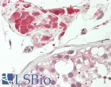
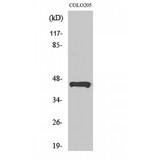
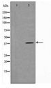
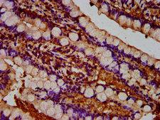
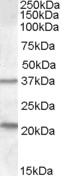
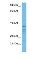
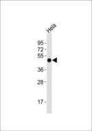
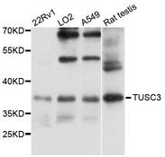
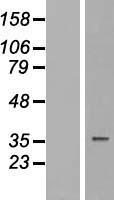
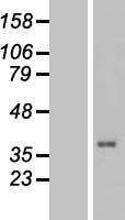
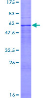
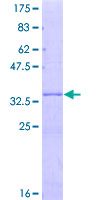
TUSC3
tumor suppressor candidate 3
TUSC3 is a candidate tumor suppressor gene. It is located within a homozygously deleted region of a metastatic prostate cancer. The gene is expressed in most nonlymphoid human tissues including prostate, lung, liver, and colon. Expression was also detected in many epithelial tumor cell lines. Two transcript variants encoding distinct isoforms have been identified for this gene.
| Gene Name: | tumor suppressor candidate 3 |
| Synonyms: | TUSC3, D8S1992, M33, MRT7, N33, OST3A, Tumor suppressor candidate 3, Protein N33, MRT22 |
| Target Sequences: | NM_006765 NP_006756.2 Q13454 |
Publications (2)


If you do not find the reagent or information you require, please contact Customer.Support@LSBio.com to inquire about additional products in development.


