order histories, retained contact details for faster checkout, review submissions, and special promotions.
Forgot password?
order histories, retained contact details for faster checkout, review submissions, and special promotions.
Locations
Orders Processing,
Shipping & Receiving,
Warehouse
2 Shaker Rd Suites
B001/B101
Shirley, MA 01464
Production Lab
Floor 6, Suite 620
20700 44th Avenue W
Lynnwood, WA 98036
Telephone Numbers
Tel: +1 (206) 374-1102
Fax: +1 (206) 577-4565
Contact Us
Additional Contact Details
order histories, retained contact details for faster checkout, review submissions, and special promotions.
Forgot password?
order histories, retained contact details for faster checkout, review submissions, and special promotions.
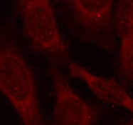
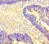
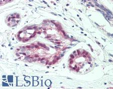
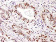
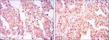
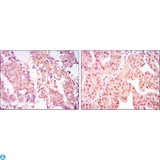
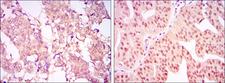
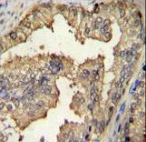
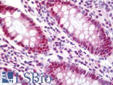
PRMT4 / CARM1
coactivator-associated arginine methyltransferase 1
Methylates (mono- and asymmetric dimethylation) the guanidino nitrogens of arginyl residues in several proteins involved in DNA packaging, transcription regulation, pre-mRNA splicing, and mRNA stability. Recruited to promoters upon gene activation together with histone acetyltransferases from EP300/P300 and p160 families, methylates histone H3 at 'Arg-17' (H3R17me), forming mainly asymmetric dimethylarginine (H3R17me2a), leading to activate transcription via chromatin remodeling. During nuclear hormone receptor activation and TCF7L2/TCF4 activation, acts synergically with EP300/P300 and either one of the p160 histone acetyltransferases NCOA1/SRC1, NCOA2/GRIP1 and NCOA3/ACTR or CTNNB1/beta-catenin to activate transcription. During myogenic transcriptional activation, acts together with NCOA3/ACTR as a coactivator for MEF2C. During monocyte inflammatory stimulation, acts together with EP300/P300 as a coactivator for NF-kappa-B. Acts as coactivator for PPARG, promotes adipocyte differentiation and the accumulation of brown fat tissue. Plays a role in the regulation of pre-mRNA alternative splicing by methylation of splicing factors. Also seems to be involved in p53/TP53 transcriptional activation. Methylates EP300/P300, both at 'Arg-2142', which may loosen its interaction with NCOA2/GRIP1, and at 'Arg-580' and 'Arg-604' in the KIX domain, which impairs its interaction with CREB and inhibits CREB-dependent transcriptional activation. Also methylates arginine residues in RNA-binding proteins PABPC1, ELAVL1 and ELAV4, which may affect their mRNA-stabilizing properties and the half-life of their target mRNAs.
| Gene Name: | coactivator-associated arginine methyltransferase 1 |
| Synonyms: | CARM1, PRMT4 |
| Target Sequences: | NM_199141 NP_954592.1 Q86X55 |

If you do not find the reagent or information you require, please contact Customer.Support@LSBio.com to inquire about additional products in development.


