Login
Registration enables users to use special features of this website, such as past
order histories, retained contact details for faster checkout, review submissions, and special promotions.
order histories, retained contact details for faster checkout, review submissions, and special promotions.
Forgot password?
Registration enables users to use special features of this website, such as past
order histories, retained contact details for faster checkout, review submissions, and special promotions.
order histories, retained contact details for faster checkout, review submissions, and special promotions.
Quick Order
Products
Antibodies
ELISA and Assay Kits
Research Areas
Infectious Disease
Resources
Purchasing
Reference Material
Contact Us
Locations
Orders Processing,
Shipping & Receiving,
Warehouse
2 Shaker Rd Suites
B001/B101
Shirley, MA 01464
Production Lab
Floor 6, Suite 620
20700 44th Avenue W
Lynnwood, WA 98036
Telephone Numbers
Tel: +1 (206) 374-1102
Fax: +1 (206) 577-4565
Contact Us
Additional Contact Details
Login
Registration enables users to use special features of this website, such as past
order histories, retained contact details for faster checkout, review submissions, and special promotions.
order histories, retained contact details for faster checkout, review submissions, and special promotions.
Forgot password?
Registration enables users to use special features of this website, such as past
order histories, retained contact details for faster checkout, review submissions, and special promotions.
order histories, retained contact details for faster checkout, review submissions, and special promotions.
Quick Order
| Catalog Number | Size | Price |
|---|---|---|
| LS-C73395-500 | 500 µg | $866 |
Monoclonal Mouse anti‑Mouse NeuN Antibody (clone A60, Biotin, IHC, WB) LS‑C73395
Monoclonal Mouse anti‑Mouse NeuN Antibody (clone A60, Biotin, IHC, WB) LS‑C73395
Antibody:
NeuN Mouse anti-Mouse Monoclonal (Biotin) (A60) Antibody
Application:
IHC, IHC-P, ICC, WB
Reactivity:
Mouse, Rat
Format:
Biotin, Unmodified
Other formats:
Toll Free North America
 206-374-1102
For Research Use Only
Overview
Antibody:
NeuN Mouse anti-Mouse Monoclonal (Biotin) (A60) Antibody
Application:
IHC, IHC-P, ICC, WB
Reactivity:
Mouse, Rat
Format:
Biotin, Unmodified
Other formats:
Specifications
Description
NeuN antibody LS-C73395 is a biotin-conjugated mouse monoclonal antibody to NeuN from mouse. It is reactive with mouse and rat. Validated for ICC, IHC and WB.
Host
Mouse
Reactivity
Mouse, Rat
(tested or 100% immunogen sequence identity)
Clonality
IgG1
Monoclonal
Clone
A60
Conjugations
Biotin.
Also available Unconjugated.
Purification
Protein A purified
Modifications
Unmodified
Immunogen
Purified cell nuclei from mouse brain.
Specificity
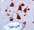
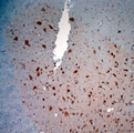
Reacts with most neuronal cell types throughout the nervous system of mice including cerebellum, cerebral cortex, hippocampus, thalamus, spinal cord and neurons in the peripheral nervous system including dorsal root ganglia, sympathetic chain ganglia and enteric ganglia. Immunohistochemical staining is primarily in the nucleus of the neurons with lighter staining in the cytoplasm. The few cell types not reactive include Purkinje, mitral and photoreceptor cells. Species cross-reactivity: Rat. It is expected that it will also react with human, ferret, chick and salamander.
Applications
- IHC
- IHC - Paraffin
- ICC (1:10 - 1:500)
- Western blot
- Applications tested for the base form of this product only

|
Performing IHC? See our complete line of Immunohistochemistry Reagents including antigen retrieval solutions, blocking agents
ABC Detection Kits and polymers, biotinylated secondary antibodies, substrates and more.
|
Usage
Suitable for use in Immunohistochemistry, Western Blot and Immunocytochemistry. Immunohistochemistry: 1:100-1:2000; Works best on polyester wax embedded tissue but also works on paraffin embedded tissue at a lower working dilution. Works well with formaldehyde-based fixatives. Citric acid and microwave pretreatment has been used successfully. Staining is primarily in the nucleus of the neurons with lighter staining in the cytoplasm. Developmentally, immunoreactivity is first observed shortly after neurons have become postmitotic, no staining has been observed in proliferative zones. Western Blot: Recognizes 2-3 bands in the 46-48kD range and possibly another band at ~66kD. Immunocytochemistry: 1:10-1:500 dilution with buffer without excess protein blocks or detergents. Neurons in culture should be permeabilized with 0.1% triton X-100. The applications listed have been tested for the unconjugated form of this product. Other forms have not been tested.
Presentation
PBS, pH 7.1, 0.09% sodium azide, 15 mg/ml BSA.
Storage
Short term: store at 4°C. Long term: aliquot and store at -20°C. Avoid freeze-thaw cycles.
Restrictions
For research use only. Intended for use by laboratory professionals.
Publications (0)
Customer Reviews (0)
Featured Products
Species:
Mouse, Human, Rat, Ferret, Chicken
Applications:
ICC, Western blot

Species:
Mouse
Applications:
IHC - Paraffin, IHC - Frozen
Request SDS/MSDS
To request an SDS/MSDS form for this product, please contact our Technical Support department at:
Technical.Support@LSBio.com
Requested From: United States
Date Requested: 4/26/2024
Date Requested: 4/26/2024


