Login
Registration enables users to use special features of this website, such as past
order histories, retained contact details for faster checkout, review submissions, and special promotions.
order histories, retained contact details for faster checkout, review submissions, and special promotions.
Forgot password?
Registration enables users to use special features of this website, such as past
order histories, retained contact details for faster checkout, review submissions, and special promotions.
order histories, retained contact details for faster checkout, review submissions, and special promotions.
Quick Order
Products
Antibodies
ELISA and Assay Kits
Research Areas
Infectious Disease
Resources
Purchasing
Reference Material
Contact Us
Locations
Orders Processing,
Shipping & Receiving,
Warehouse
2 Shaker Rd Suites
B001/B101
Shirley, MA 01464
Production Lab
Floor 6, Suite 620
20700 44th Avenue W
Lynnwood, WA 98036
Telephone Numbers
Tel: +1 (206) 374-1102
Fax: +1 (206) 577-4565
Contact Us
Additional Contact Details
Login
Registration enables users to use special features of this website, such as past
order histories, retained contact details for faster checkout, review submissions, and special promotions.
order histories, retained contact details for faster checkout, review submissions, and special promotions.
Forgot password?
Registration enables users to use special features of this website, such as past
order histories, retained contact details for faster checkout, review submissions, and special promotions.
order histories, retained contact details for faster checkout, review submissions, and special promotions.
Quick Order
| Catalog Number | Size | Price |
|---|---|---|
| LS-C204550-100 | 100 µl | $376 |
1 of 2
2 of 2
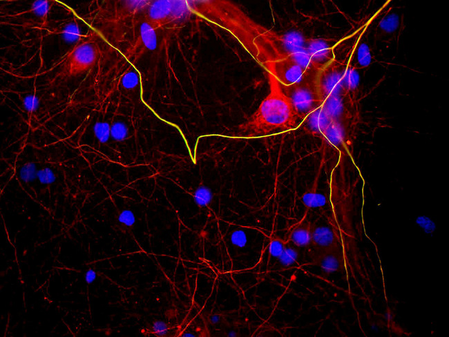
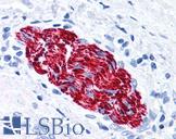
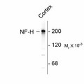
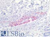
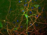
Monoclonal Mouse anti‑Bovine NEFH / NF‑H Antibody (clone AH1, IF, WB) LS‑C204550
Monoclonal Mouse anti‑Bovine NEFH / NF‑H Antibody (clone AH1, IF, WB) LS‑C204550
Antibody:
NEFH / NF-H Mouse anti-Bovine Monoclonal (AH1) Antibody
Application:
ICC, IF, WB
Reactivity:
Bovine, Chicken, Mammal
Format:
Unconjugated, Unmodified
Toll Free North America
 206-374-1102
For Research Use Only
Overview
Antibody:
NEFH / NF-H Mouse anti-Bovine Monoclonal (AH1) Antibody
Application:
ICC, IF, WB
Reactivity:
Bovine, Chicken, Mammal
Format:
Unconjugated, Unmodified
Specifications
Description
NF-H antibody LS-C204550 is an unconjugated mouse monoclonal antibody to NF-H (NEFH) from bovine. It is reactive with chicken and mammal. Validated for ICC, IF and WB.
Target
Bovine NEFH / NF-H
Synonyms
NEFH | 200 kDa neurofilament protein | KIAA0845 | NFH | NF-H
Host
Mouse
Reactivity
Bovine, Chicken, Mammal
(tested or 100% immunogen sequence identity)
Clonality
IgG1,k
Monoclonal
Clone
AH1
Conjugations
Unconjugated
Purification
Ascites
Modifications
Unmodified
Immunogen
Native NF-H purified from bovine spinal cord.
Specificity
Clone AH1 recognizes phosphorylated NF-H KSP (lysine-serine-proline) type sequences. In some species there is some cross-reactivity with the related phosphorylated KSP sequences found in the related neurofilament subunit NF-M. The antibody recognizes NF-H strongly in all mammals tested to date and also in chicken. It recognizes neurofilaments in frozen sections in tissue culture and in formalin fixed sections.
Applications
- ICC (1:1000)
- Immunofluorescence (1:1000)
- Western blot (1:10000)
Usage
The ascites solution has a high titre and can be used at dilutions of at least 1:1000 in immunofluorescence experiments. In western blotting using chemiluminescence it can be used at dilutions of 1:10000 or lower.
Presentation
Ascites fluid.
Storage
Store at 4°C or -20°C. Avoid freeze-thaw cycles.
Restrictions
For research use only. Intended for use by laboratory professionals.
About NEFH / NF-H
Publications (0)
Customer Reviews (0)
Featured Products
Species:
Human, Rat, Porcine
Applications:
IHC, IHC - Paraffin, IHC - Frozen, Western blot, ELISA
Species:
Bovine, Chicken, Mammal
Applications:
Immunofluorescence, Western blot
Species:
Bovine, Human, Mouse, Rat, Chicken
Applications:
ICC, Immunofluorescence, Western blot
Species:
Human
Applications:
IHC, Immunofluorescence, Western blot, ELISA
Request SDS/MSDS
To request an SDS/MSDS form for this product, please contact our Technical Support department at:
Technical.Support@LSBio.com
Requested From: United States
Date Requested: 4/25/2024
Date Requested: 4/25/2024


