Login
Registration enables users to use special features of this website, such as past
order histories, retained contact details for faster checkout, review submissions, and special promotions.
order histories, retained contact details for faster checkout, review submissions, and special promotions.
Forgot password?
Registration enables users to use special features of this website, such as past
order histories, retained contact details for faster checkout, review submissions, and special promotions.
order histories, retained contact details for faster checkout, review submissions, and special promotions.
Quick Order
Products
Antibodies
ELISA and Assay Kits
Research Areas
Infectious Disease
Resources
Purchasing
Reference Material
Contact Us
Locations
Orders Processing,
Shipping & Receiving,
Warehouse
2 Shaker Rd Suites
B001/B101
Shirley, MA 01464
Production Lab
Floor 6, Suite 620
20700 44th Avenue W
Lynnwood, WA 98036
Telephone Numbers
Tel: +1 (206) 374-1102
Fax: +1 (206) 577-4565
Contact Us
Additional Contact Details
Login
Registration enables users to use special features of this website, such as past
order histories, retained contact details for faster checkout, review submissions, and special promotions.
order histories, retained contact details for faster checkout, review submissions, and special promotions.
Forgot password?
Registration enables users to use special features of this website, such as past
order histories, retained contact details for faster checkout, review submissions, and special promotions.
order histories, retained contact details for faster checkout, review submissions, and special promotions.
Quick Order
| Catalog Number | Size | Price |
|---|---|---|
| LS-C19041-100 | 100 µg (1.67 mg/ml) | $567 |
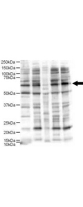
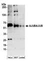
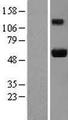
![JUB / Ajuba Antibody - Anti-Ajuba Antibody - Western Blot. Western blot of Affinity Purified anti-Ajuba antibody shows detection of Ajuba-RFP fusion protein in cell lysates (arrow-head). Lanes correspond to 1) vector only transfection, 2) human Ajuba-RFP, 3) mouse Ajuba-RFP, and 4) mock transfection. Approximately 50 ug of each lysate was loaded per lane for SDS-PAGE followed by transfer onto nitrocellulose and reaction with a 1:1700 dilution of anti-Ajuba antibody. Detection occurred using a 1:10000 dilution of IRDye800 conjugated Gt-a-Rabbit IgG [H&L] ( for 45 min at room temperature (800 nm channel, green). Molecular weight estimation was made by comparison to prestained MW markers (indicated at left, 700 nm channel, red). IRDye800 fluorescence image was captured using the Odyssey Infrared Imaging System developed by LI-COR. IRDye is a trademark of LI-COR, Inc. Other detection systems will yield similar results.](https://lsbio-7d62.kxcdn.com/image2/jub-ajuba-antibody-ls-c19041/62891_51408.jpg)
1 of 2
2 of 2
Polyclonal Rabbit anti‑Human JUB / Ajuba Antibody (WB) LS‑C19041
Polyclonal Rabbit anti‑Human JUB / Ajuba Antibody (WB) LS‑C19041
Antibody:
JUB / Ajuba Rabbit anti-Human Polyclonal Antibody
Application:
WB, IP, ELISA
Reactivity:
Human
Format:
Unconjugated, Unmodified
Toll Free North America
 206-374-1102
For Research Use Only
Overview
Antibody:
JUB / Ajuba Rabbit anti-Human Polyclonal Antibody
Application:
WB, IP, ELISA
Reactivity:
Human
Format:
Unconjugated, Unmodified
Specifications
Description
Ajuba antibody LS-C19041 is an unconjugated rabbit polyclonal antibody to human Ajuba (JUB). Validated for ELISA, IP and WB.
Target
Human JUB / Ajuba
Synonyms
AJUBA | Ajuba LIM protein | Jub, ajuba homolog | JUB | Protein ajuba
Host
Rabbit
Reactivity
Human
(tested or 100% immunogen sequence identity)
Predicted
Chimpanzee, Mouse, Rat, Dog (at least 90% immunogen sequence identity)
Clonality
IgG
Polyclonal
Conjugations
Unconjugated
Purification
Affinity purified
Modifications
Unmodified
Immunogen
This affinity purified antibody was prepared from whole rabbit serum produced by repeated immunizations with a synthetic peptide corresponding aa 224-239 of Human Ajuba.
Specificity
A BLAST analysis was used to suggest reactivity with this protein from human, rat, dog, mouse and chimpanzee based on 100% homology for the immunogen sequence. Cross reactivity with Ajuba protein homologues from other sources has not been determined.
Applications
- Western blot (1:500 - 1:2500)
- Immunoprecipitation (1:100)
- ELISA (1:20000 - 1:80000)
Usage
This affinity purified antibody has been tested for use in ELISA and by western blot. Specific conditions for reactivity should be optimized by the end user. Expect a band approximately 57 kD in size corresponding to Ajuba by western blotting in the appropriate cell lysate or extract.
Presentation
0.02 M Potassium Phosphate, pH 7.2, 0.15 M NaCl, 0.01% Sodium Azide
Storage
Store vial at -20°C prior to opening. Dilute only prior to immediate use. For extended storage aliquot contents and freeze at -20°C or below. Avoid freeze-thaw cycles.
Restrictions
For research use only. Intended for use by laboratory professionals.
About JUB / Ajuba
Publications (0)
Customer Reviews (0)
Featured Products
Species:
Human, Monkey, Mouse, Bovine
Applications:
Western blot, Immunoprecipitation, Flow Cytometry
Species:
Human
Applications:
Western blot, Immunoprecipitation
Source:
Human
Tag:
Myc-DDK (Flag)
Source:
Human
Tag:
Myc-DDK (Flag)
Request SDS/MSDS
To request an SDS/MSDS form for this product, please contact our Technical Support department at:
Technical.Support@LSBio.com
Requested From: United States
Date Requested: 4/25/2024
Date Requested: 4/25/2024


