
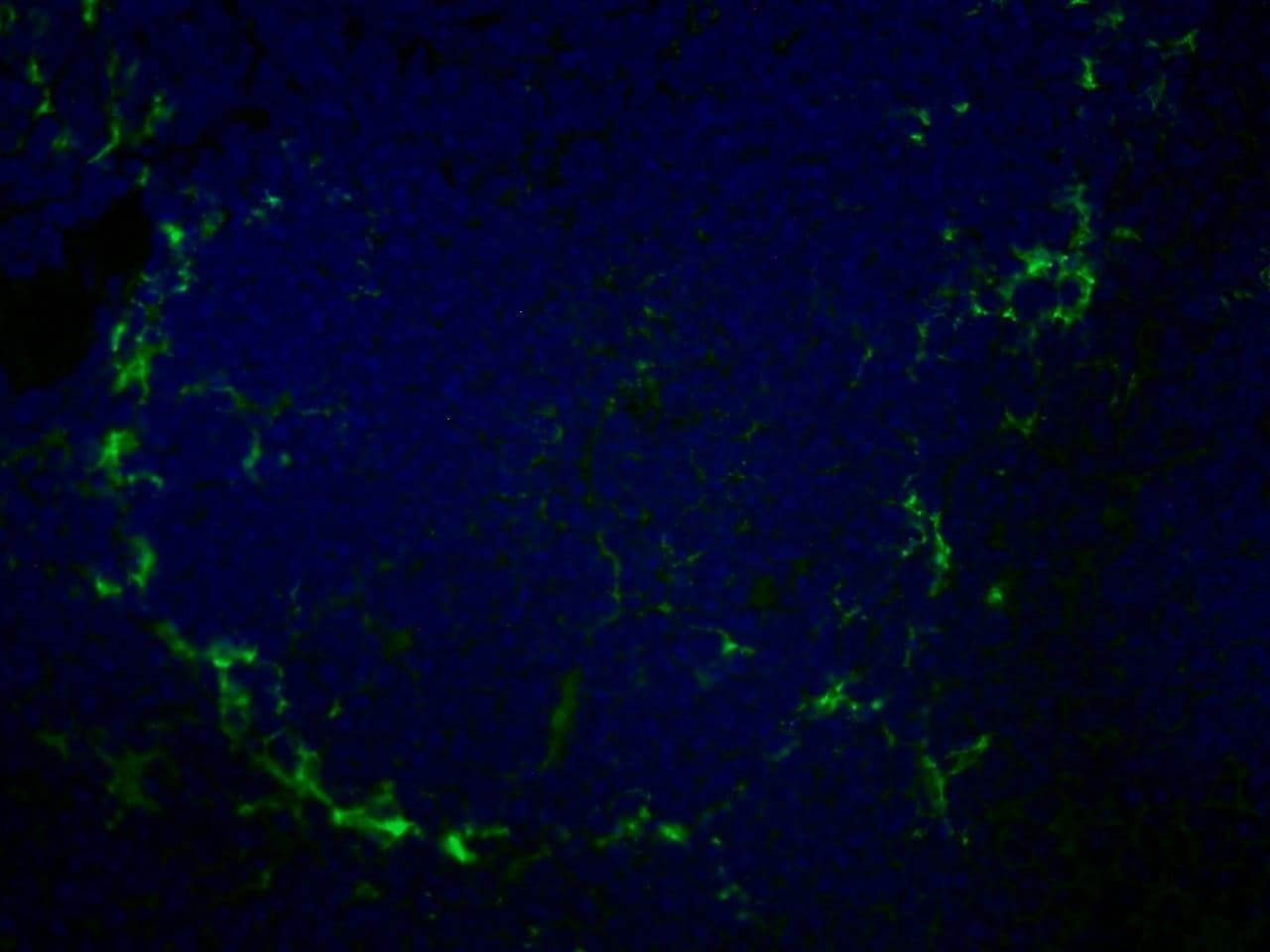
Immunohistochemistry of mouse spleen with Rat anti-Mouse CD169 This image was taken for the unconjugated form of this product. Other forms have not been tested.
SIGLEC1 / CD169 / Sialoadhesin Antibody (clone MOMA-1, FITC)
Primary AntibodyLSBio Guarantee
| Antibody: | Rat Monoclonal |
| Applications: | Flow Cytometry, Immunofluorescence, Immunohistochemistry (general) |
| Reactivity: | Mouse |
| Format: | Liquid |
$401.00each
Catalog Number: LS-C188785-0.1
Catalog Number: LS-C188785
For bulk orders or custom formulations:
Request a QuoteOverview
- Description
- Sialoadhesin antibody LS-C188785 is an FITC-conjugated rat monoclonal antibody to mouse Sialoadhesin (SIGLEC1 / CD169). Validated for Flow and IF. Cited in 1 publication.
- Application
- FC, IF, IHC
- Reactivity
- Ms
- Nonreactive Species
- Human, Rat
- Intended Use
- Research Use Only
- Guarantees
- This antibody carries the LSBio 100% Guarantee. ✓
- Category
- Antibodies > Primary Antibodies > Monoclonal Antibodies
- Alternative Names
- CD169 | CD169 antigen | SIGLEC-1 | SIGLEC1 | Sialoadhesin | SN | DJ1009E24.1
- UniProt Number
- Q9BZZ2
- NCBI GenBank
- NM_023068 | NP_075556.1
- SwissProt
- Q9BZZ2
Recommended Dilution
| Application | Dilution |
|---|---|
| FC | 0.0423611111111111 |
Target
- Target
- Mouse SIGLEC1 / CD169 / Sialoadhesin
- Antigen Species
- Mouse
- Epitope
- Specific for CD169, also known as sialoadhesin. The antibody reacts with a subpopulation of mature resident tissue macrophages. No reactivity is seen with dendritic cells, peritoneal resident macrophages, peritoneal exudate cells or blood cells. Distinct macrophage subpopulations of lymphoid organs express the antigen. In the spleen, they are localized at the marginal sinus forming a ring around the periarteriolar lymphocyte sheath and follicular areas at the inner side of marginal zones. In lymph nodes, they are localized in the sinusoids and medullary cords but not within follicular areas or paracortex. In Peyer's patches they are localized in the interfollicular areas at the serosal side. Kupffer cells in the liver can be clearly stained by MOMA-1, although antigen expression is weaker than that seen in splenic macrophages. No MOMA-1 positive macrophages were found in the thymus. Reactivity was also negative in following organs tested so far (kidney, brain, skin). In non-lymphoid organs, the antigen is only found on a macrophage subpopulation in the lamina propria of the villi of the small intestine.
- Gene Name
- SIGLEC1
Antibody
- Host Species
- Rat
- Clonality
- Monoclonal
- Clone
- MOMA-1
- Isotype
- IgG2a
Immunogen
- Immunogen
- Stromal (reticular) elements from spleen.
Format
- Conjugate/Label
- Fluorescein Isothiocyanate
- Format
- Liquid
- Formulation
- PBS, 0.09% Sodium Azide, 1% BSA
- Concentration
- 0.1 mg/mL
- Preservative
- Yes
- Purification Method
- Protein G Affinity
Storage & Handling
- Storage Conditions
- Store at 4°C or at -20°C. Store undiluted. Avoid freeze-thaw cycles. Protect from light. Microcentrifugation recommended if solution contains precipitate.
- Recommended Storage Buffer
- PBS
Pericyte MyD88 and IRAK4 control inflammatory and fibrotic responses to tissue injury. Leaf IA, Nakagawa S, Johnson BG, Cha JJ, Mittelsteadt K, Guckian KM, Gomez IG, Altemeier WA, Duffield JS. The Journal of clinical investigation. 2017;127:321-334. PubMed: 27869651 PMC: PMC5199713