LSBio
PathPlus™ TP53 / p53 Antibody (clone rBP53-12)

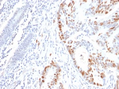
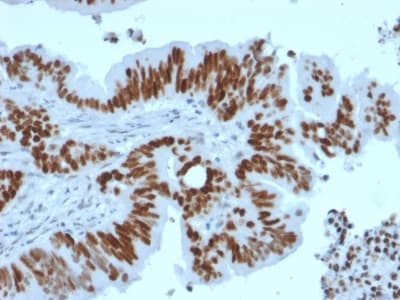
Formalin-paraffin human Colon Carcinoma stained with p53 Mouse Recombinant Monoclonal Antibody (rBP53-12).

PathPlus™ TP53 / p53 Antibody (clone rBP53-12)

Primary AntibodyLSBio Guarantee
Antibody:Mouse Recombinant Monoclonal
Applications:Immunohistochemistry (general), Immunohistochemistry, Paraffin, Western Blot
Reactivity:Human
Format:Liquid
See all TP53 / p53 antibodies →
$536.00each
Catalog Number: LS-B16261-100

For bulk orders or custom formulations:

Request a Quote

Overview

Description
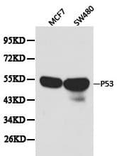
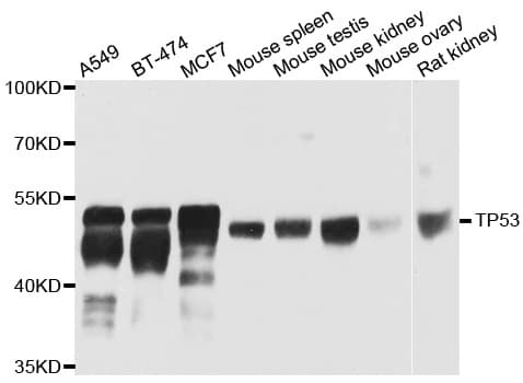
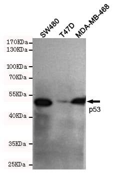
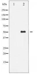
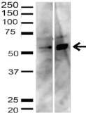
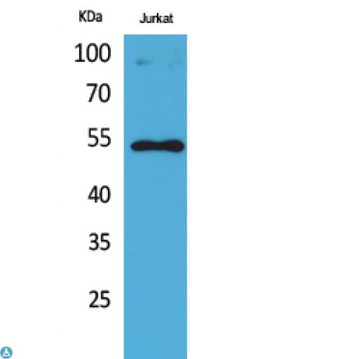
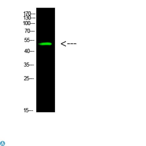
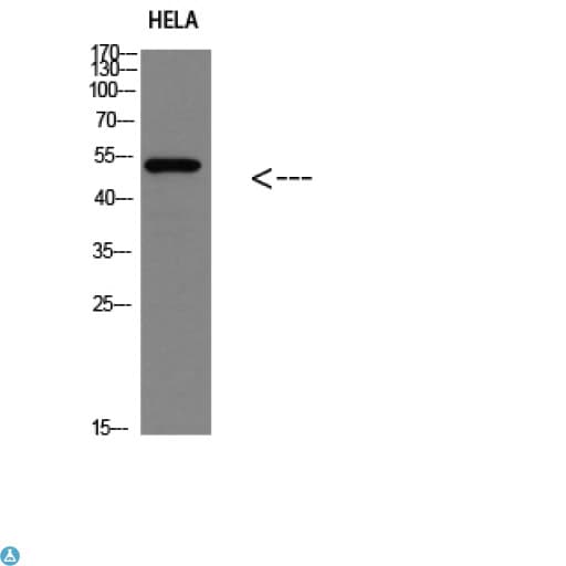
P53 antibody LS-B16261 is an unconjugated mouse recombinant monoclonal antibody to human p53 (TP53). Validated for IHC and WB.
Application
IHC, IHC-P, WB
Reactivity
Hu
Nonreactive Species
Mouse, Rat
Intended Use
Research Use Only
Guarantees
This product carries the LSBio 100% Guarantee.
Category
Antibodies > Primary Antibodies > Recombinant Antibodies
Alternative Names
BCC7 | Cellular tumor antigen p53 | p53 | TRP53 | TP53 | p53 tumor suppressor | Phosphoprotein p53 | Antigen NY-CO-13 | Tumor protein p53 | Tumor suppressor p53 | LFS1
Recommended Dilution: IHC-P
1:50
Recommended Dilution: WB
0.5 - 1 µg/ml
UniProt Number
P04637
SwissProt
P04637

Target

Target
TP53 / p53
Antigen Species
Human
Specificity
This MAb reacts with an N-terminal epitope (aa 16-25) of both wild type and mutated p53. Mutation and/or allelic loss of p53 is one of the causes of a variety of mesenchymal and epithelial tumors. If it occurs in the germ line, such tumors run in families. In most transformed and tumor cells the concentration of p53 is increased 51000 fold over the minute concentrations (1000 molecules cell) in normal cells, principally due to the increased half-life (4 h) compared to that of the wild-type (20 min). p53 Localizes in the nucleus, but is detectable at the plasma membrane during mitosis and when certain mutations modulate cytoplasmic/nuclear distribution. Mutations arise with an average frequency of 70% but incidence varies from zero in carcinoid lung tumors to 97% in primary melanomas. High concentrations of p53 protein are transiently expressed in human epidermis and superficial dermal fibroblasts following mild ultraviolet irradiation. Positive nuclear staining with p53 antibody has been reported to be a negative prognostic factor in breast carcinoma, lung carcinoma, colorectal, and urothelial carcinoma. Anti-p53 positivity has also been used to differentiate uterine serous carcinoma from endometrioid carcinoma as well as to detect intratubular germ cell neoplasia.
Epitope
This MAb reacts with an N-terminal epitope of both wild type and mutated p53. Mutation and/or allelic loss of p53 is one of the causes of a variety of mesenchymal and epithelial tumors. If it occurs in the germ line, such tumors run in families. In most transformed and tumor cells the concentration of p53 is increased 51000 fold over the minute concentrations (1000 molecules cell) in normal cells, principally due to the increased half-life (4 h) compared to that of the wild-type (20 min). p53 Localizes in the nucleus, but is detectable at the plasma membrane during mitosis and when certain mutations modulate cytoplasmic/nuclear distribution. Mutations arise with an average frequency of 70% but incidence varies from zero in carcinoid lung tumors to 97% in primary melanomas. High concentrations of p53 protein are transiently expressed in human epidermis and superficial dermal fibroblasts following mild ultraviolet irradiation. Positive nuclear staining with p53 antibody has been reported to be a negative prognostic factor in breast carcinoma, lung carcinoma, colorectal, and urothelial carcinoma. Anti-p53 positivity has also been used to differentiate uterine serous carcinoma from endometrioid carcinoma as well as to detect intratubular germ cell neoplasia.
Gene Name
TP53

Antibody

Host Species
Mouse
Clonality
Recombinant Monoclonal
Clone
rBP53-12
Origin Species
Human

Immunogen

Immunogen
Recombinant human wild-type p53 protein. Antigen Molecular Weight: 53 kDa

Format

Conjugate/Label
Unconjugated
Format
Liquid
Formulation
10 mM PBS, 0.05% BSA, 0.05% Sodium Azide
Concentration
200 µg/mL
Animal Free
No
Carrier-Free
No
Preservative
Yes
Purification Method
Protein A/G Affinity

Storage & Handling

Storage Conditions
Store at 2°C to 8°C for up to 2 years.
Recommended Storage Buffer
10 mM PBS

Request SDS/MSDS

To request an SDS/MSDS form for this product, please contact our Technical Support department at: technical@vectorlabs.com.