
Melanoma Associated Antigen KBA.62 Antibody (Concentrated)
Catalog Number: LS-C761963
2 sizes / from $277.00
Antibody: Mouse Monoclonal
Applications: Enzyme-Linked Immunosorbent Assay, Immunohistochemistry (general)
Reactivity: Human
Format: Liquid

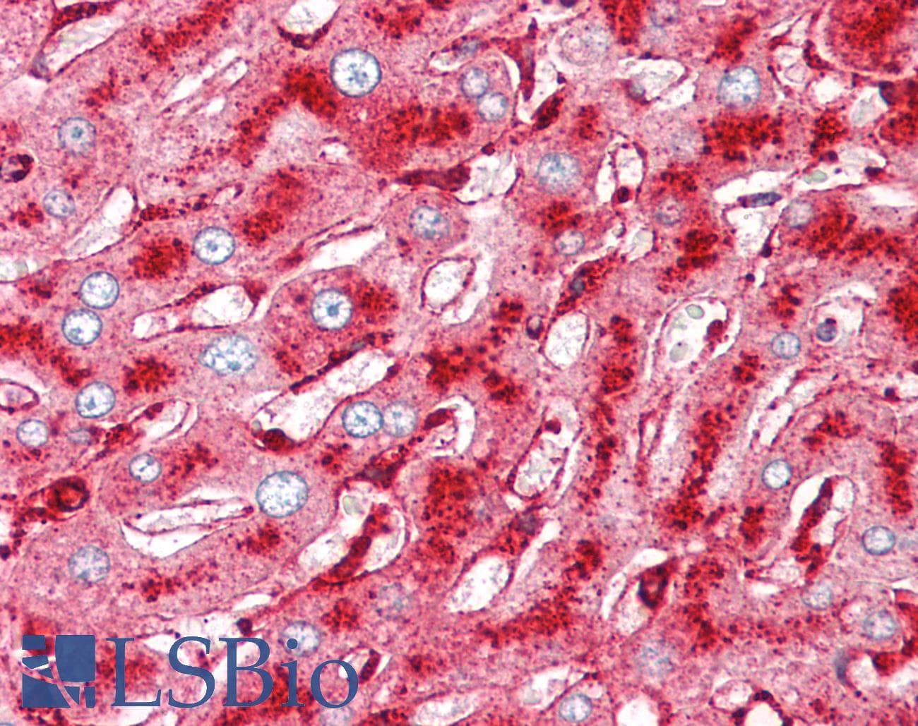
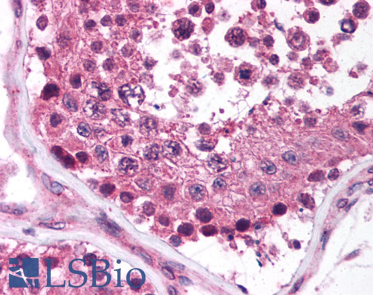
Anti-Melanoma-Associated Antigen antibody IHC of human liver. Immunohistochemistry of formalin-fixed, paraffin-embedded tissue after heat-induced antigen retrieval. Antibody concentration 10 ug/ml.
| Antibody: | Mouse Monoclonal |
| Applications: | Enzyme-Linked Immunosorbent Assay, Immunofluorescence, Immunohistochemistry (general), Immunohistochemistry, Frozen, Immunohistochemistry, Paraffin, Immunoprecipitation |
| Reactivity: | Human |
| Format: | Liquid |
For bulk orders or custom formulations:
Request a QuoteCatalog Number: LS-C761963
2 sizes / from $277.00
Antibody: Mouse Monoclonal
Applications: Enzyme-Linked Immunosorbent Assay, Immunohistochemistry (general)
Reactivity: Human
Format: Liquid
Catalog Number: LS-C683678-0.1
0.1 mL / $309.00
Antibody: Mouse Monoclonal
Applications: Immunohistochemistry (general)
Reactivity: Human
Format: Liquid
Catalog Number: LS-C783586-100
100 µL / $394.00
Antibody: Mouse Monoclonal
Applications: Immunohistochemistry (general)
Reactivity: Human
Format: Liquid
Catalog Number: LS-C761964-7
7 mL / $378.00
Antibody: Mouse Monoclonal
Applications: Enzyme-Linked Immunosorbent Assay, Immunohistochemistry (general)
Reactivity: Human
Format: Liquid
Catalog Number: LS-C202878-1
1 mL / $1113.00
Antibody: Mouse Monoclonal
Applications: Immunohistochemistry (general), Immunohistochemistry, Frozen, Immunohistochemistry, Paraffin
Reactivity: Human
Format: Liquid

Catalog Number: LS-F12992-1
1 plate / $683.00
Reactivity: Human
Detection Method Colorimetric
Format: 96-Well Strip Plate

Catalog Number: LS-F20102-1
1 plate / $683.00
Reactivity: Mouse
Detection Method Colorimetric
Format: 96-Well Strip Plate
Catalog Number: LS-F35168-1
1 plate / $591.00
Reactivity: Human
Detection Method Colorimetric
Format: 96-Well Strip Plate
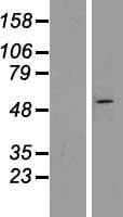
Catalog Number: LS-G111818-100
100 µg (1 mg/mL) / $725.00
Expression System Human Embryonic Kidney 293
Format: Liquid
Catalog Number: LS-G86881-20
20 µg / $220.00
Expression System Human Embryonic Kidney 293T
Format: Lyophilized
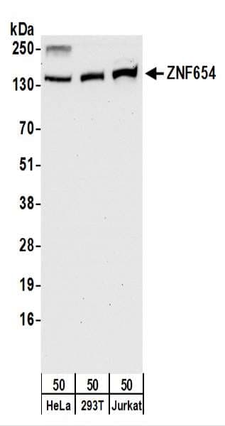
Catalog Number: LS-C290989
2 sizes / from $301.00
Antibody: Rabbit Polyclonal
Applications: Immunoprecipitation, Western Blot
Reactivity: Human
Format: Liquid
Catalog Number: LS-C660796-100
100 µg / $359.00
Antibody: Rabbit Polyclonal
Applications: Enzyme-Linked Immunosorbent Assay, Immunofluorescence, Immunohistochemistry (general), Western Blot
Reactivity: Human
Format: Liquid
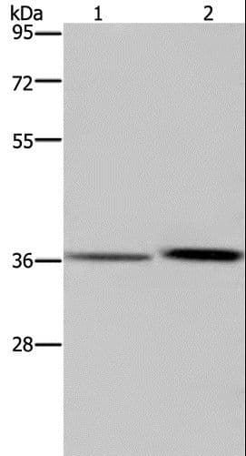
Catalog Number: LS-C403314
4 sizes / from $260.00
Antibody: Rabbit Polyclonal
Applications: Enzyme-Linked Immunosorbent Assay, Immunohistochemistry (general), Western Blot
Reactivity: Human
Format: Liquid
Catalog Number: LS-C666609-100
100 µg / $359.00
Antibody: Rabbit Polyclonal
Applications: Enzyme-Linked Immunosorbent Assay, Immunohistochemistry (general), Western Blot
Reactivity: Human
Format: Liquid
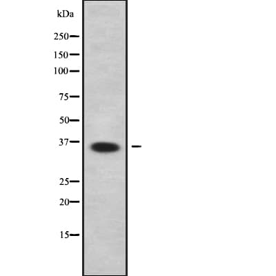
Catalog Number: LS-C807551
2 sizes / from $288.00
Antibody: Rabbit Polyclonal
Applications: Enzyme-Linked Immunosorbent Assay, Immunohistochemistry (general), Western Blot
Reactivity: Human, Mouse
Format: Liquid
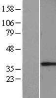
Catalog Number: LS-G110424-100
100 µg (1 mg/mL) / $504.00
Expression System Human Embryonic Kidney 293
Format: Liquid
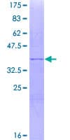
Catalog Number: LS-G63069
2 sizes / from $489.00
Expression System Tobacco / Plant-Based Expression
Format: Liquid
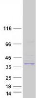
Catalog Number: LS-G72173-20
20 µg / $1130.00
Expression System Human Embryonic Kidney 293
Format: Liquid
Catalog Number: LS-G91604-20
20 µg / $153.00
Expression System Human Embryonic Kidney 293T
Format: Lyophilized
Catalog Number: LS-B893-50
50 µg (1 mg/mL) / $383.00
Antibody: Rabbit Polyclonal
Applications: Enzyme-Linked Immunosorbent Assay, Immunohistochemistry (general), Immunohistochemistry, Paraffin
Reactivity: Human
Format: Liquid