Login
Registration enables users to use special features of this website, such as past
order histories, retained contact details for faster checkout, review submissions, and special promotions.
order histories, retained contact details for faster checkout, review submissions, and special promotions.
Forgot password?
Registration enables users to use special features of this website, such as past
order histories, retained contact details for faster checkout, review submissions, and special promotions.
order histories, retained contact details for faster checkout, review submissions, and special promotions.
Quick Order
Products
Antibodies
ELISA and Assay Kits
Research Areas
Infectious Disease
Resources
Purchasing
Reference Material
Contact Us
Locations
Orders Processing,
Shipping & Receiving,
Warehouse
2 Shaker Rd Suites
B001/B101
Shirley, MA 01464
Production Lab
Floor 6, Suite 620
20700 44th Avenue W
Lynnwood, WA 98036
Telephone Numbers
Tel: +1 (206) 374-1102
Fax: +1 (206) 577-4565
Contact Us
Additional Contact Details
Login
Registration enables users to use special features of this website, such as past
order histories, retained contact details for faster checkout, review submissions, and special promotions.
order histories, retained contact details for faster checkout, review submissions, and special promotions.
Forgot password?
Registration enables users to use special features of this website, such as past
order histories, retained contact details for faster checkout, review submissions, and special promotions.
order histories, retained contact details for faster checkout, review submissions, and special promotions.
Quick Order
| Catalog Number | Size | Price |
|---|---|---|
| LS-C797459-100 | 100 µg | $511 |

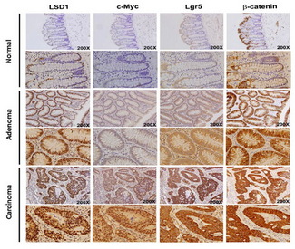

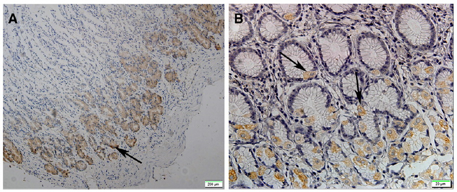
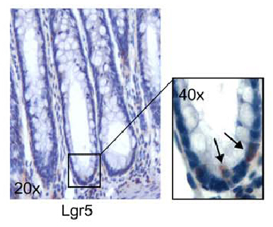


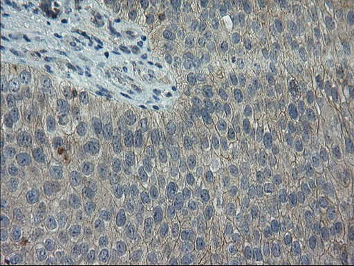

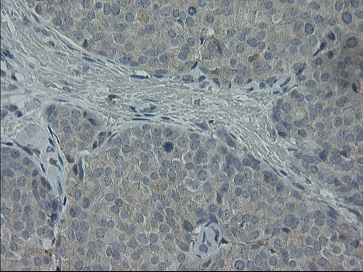
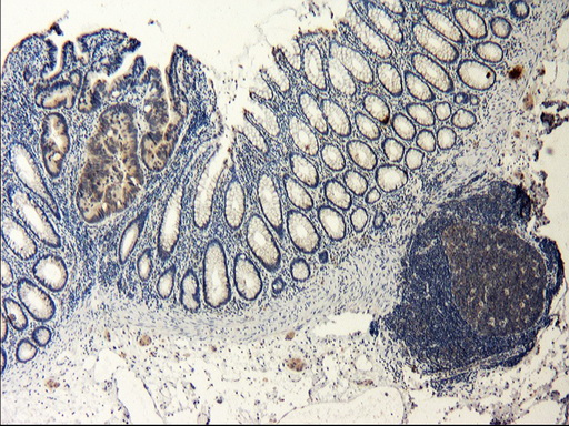


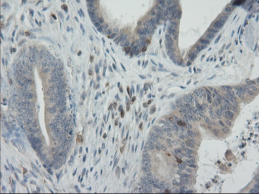

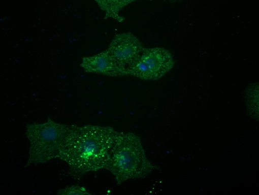
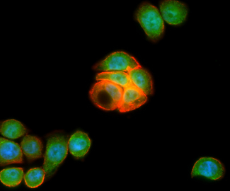
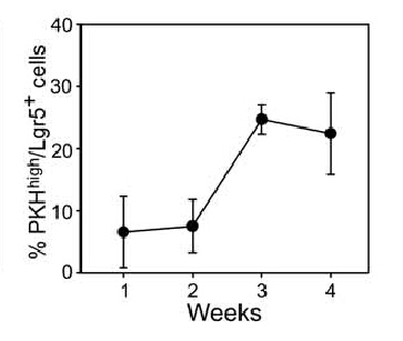
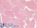
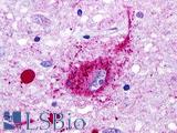
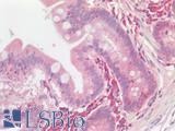
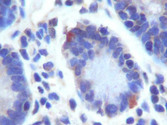
![GPR49 / LGR5 Antibody - Figure from citation: [Nature] Cells expressing the stem cell marker Lgr5 was detected by immunostaining of hCECs in 2D culture. <br />Pubmed id: 27398792<br />Memo: Human; IF](https://lsbio-7d62.kxcdn.com/image2/gpr49-lgr5-antibody-aa250-550-clone-oti2a2-carrier-free-ls-c797459/522421_4344918.jpg)
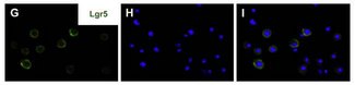

![GPR49 / LGR5 Antibody - Figure from citation: [Nature] Cells expressing the stem cell marker Lgr5 was detected by immunostaining of hCECs in 2D culture. <br />Pubmed id: 27398792<br />Memo: Human; IF](https://lsbio-7d62.kxcdn.com/image2/gpr49-lgr5-antibody-aa250-550-clone-oti2a2-carrier-free-ls-c797459/522422_4344960.jpg)


1 of 36
2 of 36
3 of 36
4 of 36
5 of 36
6 of 36
7 of 36
8 of 36
9 of 36
10 of 36
11 of 36
12 of 36
13 of 36
14 of 36
15 of 36
16 of 36
17 of 36
18 of 36
19 of 36
20 of 36
21 of 36
22 of 36
23 of 36
24 of 36
25 of 36
26 of 36
27 of 36
28 of 36
29 of 36
30 of 36
31 of 36
32 of 36
33 of 36
34 of 36
35 of 36
36 of 36
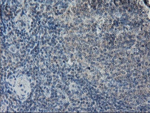
Monoclonal Mouse anti‑Human GPR49 / LGR5 Antibody (clone OTI2A2, Carrier‑free, aa250‑550, IHC, IF, WB) LS‑C797459
Monoclonal Mouse anti‑Human GPR49 / LGR5 Antibody (clone OTI2A2, Carrier‑free, aa250‑550, IHC, IF, WB) LS‑C797459
Antibody:
GPR49 / LGR5 Mouse anti-Human Monoclonal (aa250-550) (Carrier-free) (OTI2A2) Antibody
Application:
IHC, IF, WB, IP, Flo
Reactivity:
Human, Mouse
Format:
Unconjugated, Carrier-free
Other formats:
Toll Free North America
 206-374-1102
For Research Use Only
Overview
Antibody:
GPR49 / LGR5 Mouse anti-Human Monoclonal (aa250-550) (Carrier-free) (OTI2A2) Antibody
Application:
IHC, IF, WB, IP, Flo
Reactivity:
Human, Mouse
Format:
Unconjugated, Carrier-free
Other formats:
Specifications
Description
LGR5 antibody LS-C797459 is an unconjugated mouse monoclonal antibody to LGR5 (GPR49) (aa250-550) from human. It is reactive with human and mouse. Validated for Flow, IF, IHC, IP and WB.
Target
Human GPR49 / LGR5
Synonyms
LGR5 | FEX | G-protein coupled receptor 49 | G protein-coupled receptor 49 | GPR67 | GRP49 | GPR49 | HG38 | G-protein coupled receptor 67
Host
Mouse
Reactivity
Human, Mouse
(tested or 100% immunogen sequence identity)
Clonality
IgG1
Monoclonal
Clone
OTI2A2
Conjugations
Unconjugated
Purification
Purified from ascites.
Modifications
Carrier-free.
Also available Unmodified.
Immunogen
Human recombinant protein fragment corresponding to amino acids 250-550 of human LGR5 (NP_003658) produced in HEK293T Cell.
Epitope
aa250-550
Applications
- IHC (1:150)
- Immunofluorescence (1:100)
- Western blot (1:2000)
- Immunoprecipitation (4 µg/ml)
- Flow Cytometry (1:100)

|
Performing IHC? See our complete line of Immunohistochemistry Reagents including antigen retrieval solutions, blocking agents
ABC Detection Kits and polymers, biotinylated secondary antibodies, substrates and more.
|
Usage
Applications should be user optimized.
Presentation
PBS, pH 7.3, 8% Trehalose
Reconstitution
Reconstitute with PBS pH 7.3. To use this carrier-free antibody for conjugation experiment, we strongly recommend you to perform another round of desalting process.
Storage
Store at -20°C. Avoid freeze-thaw cycles.
Restrictions
For research use only. Intended for use by laboratory professionals.
About GPR49 / LGR5
Publications (0)
Customer Reviews (0)
Featured Products
Species:
Human
Applications:
IHC, IHC - Paraffin, Immunofluorescence
Species:
Human
Applications:
IHC, IHC - Paraffin
Species:
Human, Monkey, Bat
Applications:
IHC - Paraffin

Species:
Human
Applications:
IHC, IHC - Paraffin, Western blot

Species:
Human
Applications:
IHC, IHC - Paraffin, Western blot
Species:
Human
Applications:
IHC, IHC - Paraffin, Western blot
Request SDS/MSDS
To request an SDS/MSDS form for this product, please contact our Technical Support department at:
Technical.Support@LSBio.com
Requested From: United States
Date Requested: 4/28/2024
Date Requested: 4/28/2024


