Login
Registration enables users to use special features of this website, such as past
order histories, retained contact details for faster checkout, review submissions, and special promotions.
order histories, retained contact details for faster checkout, review submissions, and special promotions.
Forgot password?
Registration enables users to use special features of this website, such as past
order histories, retained contact details for faster checkout, review submissions, and special promotions.
order histories, retained contact details for faster checkout, review submissions, and special promotions.
Quick Order
Products
Antibodies
ELISA and Assay Kits
Research Areas
Infectious Disease
Resources
Purchasing
Reference Material
Contact Us
Locations
Orders Processing,
Shipping & Receiving,
Warehouse
2 Shaker Rd Suites
B001/B101
Shirley, MA 01464
Production Lab
Floor 6, Suite 620
20700 44th Avenue W
Lynnwood, WA 98036
Telephone Numbers
Tel: +1 (206) 374-1102
Fax: +1 (206) 577-4565
Contact Us
Additional Contact Details
Login
Registration enables users to use special features of this website, such as past
order histories, retained contact details for faster checkout, review submissions, and special promotions.
order histories, retained contact details for faster checkout, review submissions, and special promotions.
Forgot password?
Registration enables users to use special features of this website, such as past
order histories, retained contact details for faster checkout, review submissions, and special promotions.
order histories, retained contact details for faster checkout, review submissions, and special promotions.
Quick Order
| Catalog Number | Size | Price |
|---|---|---|
| LS-B10410-50 | 50 µl (1 mg/ml) | $460 |

1 of 3
2 of 3
3 of 3
IHC‑plus™ Monoclonal Mouse anti‑Human SAG / Arrestin Antibody (clone S128, IHC, IF, WB) LS‑B10410
IHC‑plus™ Monoclonal Mouse anti‑Human SAG / Arrestin Antibody (clone S128, IHC, IF, WB) LS‑B10410

Note: This antibody replaces LS-C204506
Antibody:
SAG / Arrestin Mouse anti-Human Monoclonal (S128) Antibody
Application:
IHC, IHC-P, IF, WB
Reactivity:
Human, Mouse, Rat, Bovine, Pig
Format:
Unconjugated, Unmodified
Toll Free North America
 206-374-1102
For Research Use Only
Overview
Antibody:
SAG / Arrestin Mouse anti-Human Monoclonal (S128) Antibody
Application:
IHC, IHC-P, IF, WB
Reactivity:
Human, Mouse, Rat, Bovine, Pig
Format:
Unconjugated, Unmodified
Specifications
Description
Arrestin antibody LS-B10410 is an unconjugated mouse monoclonal antibody to Arrestin (SAG) from human. It is reactive with human, mouse, rat and other species. Validated for IF, IHC and WB. Tested on 20 paraffin-embedded human tissues.
Target
Human SAG / Arrestin
Synonyms
SAG | 48 kDa protein | Arrestin 1 | ARRESTIN | S-arrestin | S-AG | Retinal S-antigen | Rod photoreceptor arrestin | S-antigen | RP47
Host
Mouse
Reactivity
Human, Mouse, Rat, Bovine, Pig
(tested or 100% immunogen sequence identity)
Clonality
IgG1,k
Monoclonal
Clone
S128
Conjugations
Unconjugated
Purification
Affinity purified
Modifications
Unmodified
Immunogen
Raised against recombinant bovine arrestin-1 with the first 20 amino acids of the C-Terminus truncated
Specificity
Clone S128 is known to react with visual arrestin from human, bovine, mouse, pig and rat.
Applications
- IHC
- IHC - Paraffin (1:100)
- Immunofluorescence (1:1000)
- Western blot (1:5000)

|
Performing IHC? See our complete line of Immunohistochemistry Reagents including antigen retrieval solutions, blocking agents
ABC Detection Kits and polymers, biotinylated secondary antibodies, substrates and more.
|
Usage
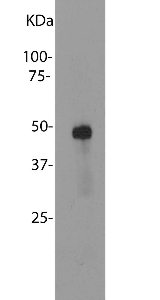
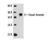
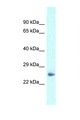
Try at dilutions of ~1:1000 for immunofluorescence. For western blots try at 1:5000. A suitable control tissue is retinal homogenate. The arrestin protein runs at about ~48 kDa on SDS-PAGE gels.
Presentation
PBS, 10 mM Sodium Azide
Storage
Store at 4°C or -20°C. Avoid freeze-thaw cycles.
Restrictions
For research use only. Intended for use by laboratory professionals.
About SAG / Arrestin
Validation
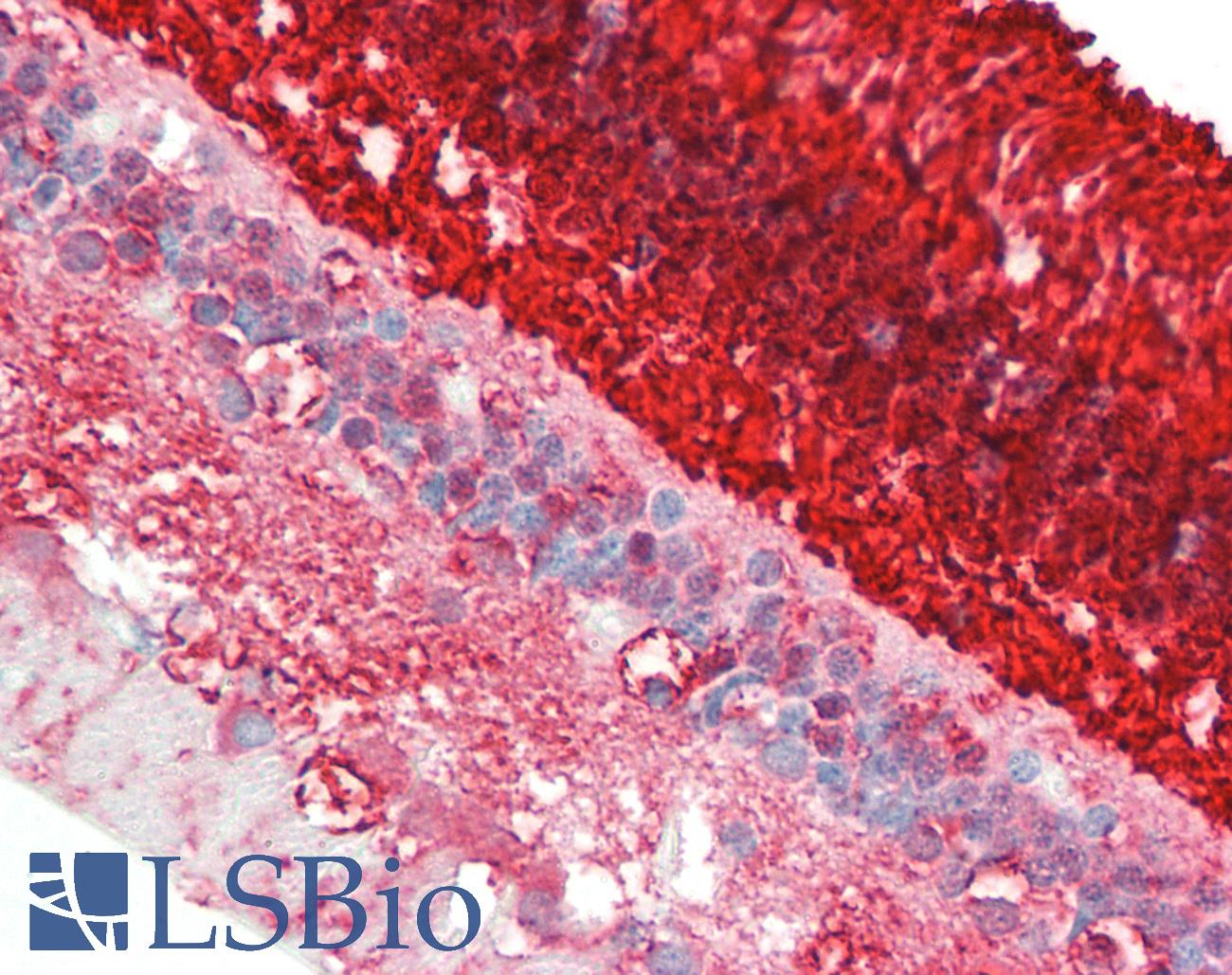
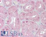
Anti-SAG / Arrestin antibody IHC staining of human retina. Immunohistochemistry of formalin-fixed, paraffin-embedded tissue after heat-induced antigen retrieval. Antibody dilution 1:100.
Anti-SAG / Arrestin antibody IHC staining of human retina. Immunohistochemistry of formalin-fixed, paraffin-embedded tissue after heat-induced antigen retrieval. Antibody dilution 1:100.
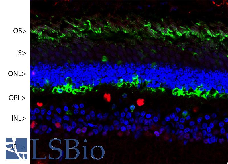
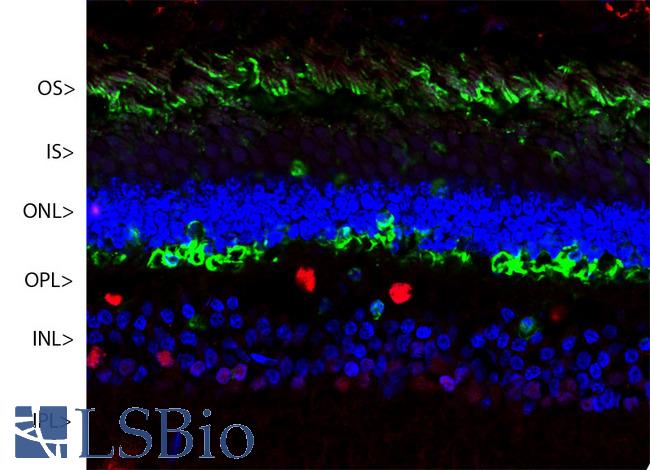
Confocal image of a pig retina stained with SAG / Arrestin antibody (green). Visual arrestin is most abundant in the outer segments (OS) and inner surface of the outer nuclear layer (ONL), and can be used to identify components of rod photoreceptor cells. (Cone photoreceptors have a different arrestin isotype). Other retinal layers are inner segments (IS), outer plexiform layer (OPL), inner nuclear layer (INL) and inner plexiform layer (IPL). The red stain is Fox2, an RNA binding nuclear protein related to Fox3/NeuN, which stains nuclei of horizontal neurons and some other neurons in the INL and IPL. Nuclear DNA was revealed with DAPI (blue).
Confocal image of a pig retina stained with SAG / Arrestin antibody (green). Visual arrestin is most abundant in the outer segments (OS) and inner surface of the outer nuclear layer (ONL), and can be used to identify components of rod photoreceptor cells. (Cone photoreceptors have a different arrestin isotype). Other retinal layers are inner segments (IS), outer plexiform layer (OPL), inner nuclear layer (INL) and inner plexiform layer (IPL). The red stain is Fox2, an RNA binding nuclear protein related to Fox3/NeuN, which stains nuclei of horizontal neurons and some other neurons in the INL and IPL. Nuclear DNA was revealed with DAPI (blue).
See More About...
LSBio Ratings
IHC-plus™ SAG / Arrestin Antibody (clone S128) for IHC, IF/Immunofluorescence, WB/Western LS-B10410 has an LSBio Rating of
Laboratory Validation Score (4)
Learn more about The LSBio Ratings Algorithm
Publications (0)
Customer Reviews (0)
Featured Products
Species:
Pig, Human, Bovine
Applications:
IHC, Western blot
Species:
Human, Bovine, Pig
Applications:
Immunofluorescence, Western blot
Reactivity:
Human
Range:
250-5000 pg/ml
Species:
Human, Mouse, Rat
Applications:
IHC, IHC - Paraffin, Peptide Enzyme-Linked Immunosorbent Assay
Request SDS/MSDS
To request an SDS/MSDS form for this product, please contact our Technical Support department at:
Technical.Support@LSBio.com
Requested From: United States
Date Requested: 4/19/2024
Date Requested: 4/19/2024


