Login
Registration enables users to use special features of this website, such as past
order histories, retained contact details for faster checkout, review submissions, and special promotions.
order histories, retained contact details for faster checkout, review submissions, and special promotions.
Forgot password?
Registration enables users to use special features of this website, such as past
order histories, retained contact details for faster checkout, review submissions, and special promotions.
order histories, retained contact details for faster checkout, review submissions, and special promotions.
Quick Order
Products
Antibodies
ELISA and Assay Kits
Research Areas
Infectious Disease
Resources
Purchasing
Reference Material
Contact Us
Locations
Orders Processing,
Shipping & Receiving,
Warehouse
2 Shaker Rd Suites
B001/B101
Shirley, MA 01464
Production Lab
Floor 6, Suite 620
20700 44th Avenue W
Lynnwood, WA 98036
Telephone Numbers
Tel: +1 (206) 374-1102
Fax: +1 (206) 577-4565
Contact Us
Additional Contact Details
Login
Registration enables users to use special features of this website, such as past
order histories, retained contact details for faster checkout, review submissions, and special promotions.
order histories, retained contact details for faster checkout, review submissions, and special promotions.
Forgot password?
Registration enables users to use special features of this website, such as past
order histories, retained contact details for faster checkout, review submissions, and special promotions.
order histories, retained contact details for faster checkout, review submissions, and special promotions.
Quick Order
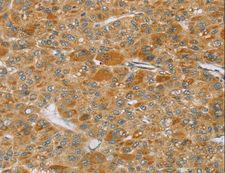
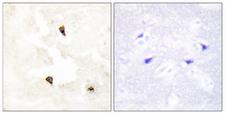
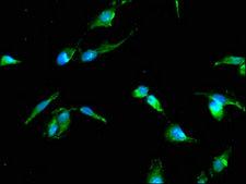
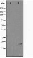
TP53I11 / PIG11
tumor protein p53 inducible protein 11
| Gene Name: | tumor protein p53 inducible protein 11 |
| Synonyms: | TP53I11, p53-induced gene 11 protein, PIG11 |
| Target Sequences: | NM_006034 NP_006025.2 O14683 |
☰ Filters
Products
Antibodies
(15)
Proteins
(6)
ELISA Kits
(1)
Type
Cell-Based
(1)
Over-Expression Lysate
(5)
Primary
(15)
Recombinant
(1)
Target
TP53I11 / PIG11
(22)
Reactivity
Human
(16)
Mouse
(3)
Rat
(2)
Application
IHC
(9)
IHC-P
(2)
WB
(9)
ELISA
(8)
ICC
(1)
IF
(4)
Peptide-ELISA
(2)
Host
rabbit
(15)
Product Group
IHCPlus
(1)
Isotype
IgG
(11)
Clonality
polyclonal pc
(15)
Format
96-Well Microplate
(1)
Biotin Conjugated
(1)
FITC Conjugated
(1)
HRP Conjugated
(1)
Unconjugated
(12)
Epitope
40-120 aa, Internal
(1)
Internal
(1)
Pro93
(1)
aa179-189
(1)
aa71-120
(1)
Publications
No
(22)
Sample Type
Adherent Cell Cultures
(1)
Tag
Myc-DDK (Flag)
(6)
Species
Human
(4)
Source
293T Cells
(3)
HEK 293 Cells
(3)
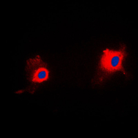
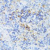
TP53I11 / PIG11 Rabbit anti-Human Polyclonal (Internal) Antibody
Human
ICC, IF, WB
Unconjugated
100 µl/$299
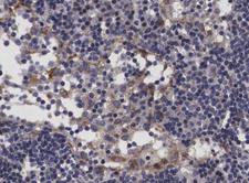
TP53I11 / PIG11 Rabbit anti-Human Polyclonal Antibody
Human
IF, IHC, Peptide-ELISA, WB
Unconjugated
100 µl/$379; 200 µl/$421
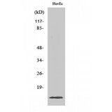
TP53I11 / PIG11 Rabbit anti-Human Polyclonal (40-120 aa, Internal) Antibody
Human
ELISA, IF, IHC, WB
Unconjugated
50 µg/$295; 100 µg/$335; 200 µg/$394
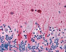

TP53I11 / PIG11 Rabbit anti-Human Polyclonal (aa179-189) Antibody
Human
ELISA, IHC, IHC-P
Unconjugated
50 µg/$460
TP53I11 / PIG11 Rabbit anti-Human Polyclonal Antibody
Mouse, Rat, Human
ELISA, IHC, WB
Unconjugated
20 µl/$254; 60 µl/$296; 120 µl/$355; 200 µl/$450
TP53I11 / PIG11 Rabbit anti-Human Polyclonal (aa71-120) Antibody
Human
IHC, IHC-P, Peptide-ELISA, WB
Unconjugated
50 µl/$334; 100 µl/$397

TP53I11 / PIG11 Rabbit anti-Human Polyclonal Antibody
Human
ELISA, IHC, WB
Unconjugated
100 µg/$357
TP53I11 / PIG11 Rabbit anti-Human Polyclonal (Pro93) Antibody
Human
IHC, WB
Unconjugated
50 µl/$321; 100 µl/$403
TP53I11 / PIG11 Rabbit anti-Human Polyclonal Antibody
Human
ELISA, IF
Unconjugated
50 µg/$294; 100 µg/$360
TP53I11 / PIG11 Rabbit anti-Human Polyclonal (HRP) Antibody
Human
ELISA
HRP Conjugated
50 µg/$294; 100 µg/$360
TP53I11 / PIG11 Rabbit anti-Human Polyclonal (Biotin) Antibody
Human
ELISA
Biotin Conjugated
50 µg/$294; 100 µg/$360
TP53I11 / PIG11 Rabbit anti-Human Polyclonal (FITC) Antibody
Human
ELISA
FITC Conjugated
50 µg/$294; 100 µg/$360
TP53I11 / PIG11 Rabbit anti-Human Polyclonal Antibody
Human
IHC, WB
Unconjugated
50 µg/$305; 100 µg/$357
TP53I11 / PIG11 Rabbit anti-Human Polyclonal Antibody
Mouse, Rat, Human
IHC
Unconjugated
60 µl/$331; 120 µl/$403; 200 µl/$529
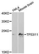
TP53I11 / PIG11 Rabbit anti-Human Polyclonal Antibody
Mouse, Human
WB
Unconjugated
20 µl/$275; 50 µl/$311; 100 µl/$389; 200 µl/$503

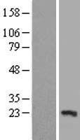
HEK 293 Cells
Myc-DDK (Flag)
20.9 kDa
100 µg/$494
HEK 293 Cells
Myc-DDK (Flag)
20.9 kDa
100 µg/$494
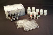
Cell-Based
96-Well Microplate
Human
Colorimetric - 450nm (TMB)
Adherent Cell Cultures
1 Plate/$618
HEK 293 Cells
Myc-DDK (Flag)
20.9 kDa
20 µg/$1,107

293T Cells
Myc-DDK (Flag)
20.9 kDa
20 µg/$150
293T Cells
Myc-DDK (Flag)
20.9 kDa
20 µg/$150

293T Cells
Myc-DDK (Flag)
20.9 kDa
20 µg/$150
Viewing 1-22
of 22
product results
If you do not find the reagent or information you require, please contact Customer.Support@LSBio.com to inquire about additional products in development.


