Login
Registration enables users to use special features of this website, such as past
order histories, retained contact details for faster checkout, review submissions, and special promotions.
order histories, retained contact details for faster checkout, review submissions, and special promotions.
Forgot password?
Registration enables users to use special features of this website, such as past
order histories, retained contact details for faster checkout, review submissions, and special promotions.
order histories, retained contact details for faster checkout, review submissions, and special promotions.
Quick Order
Products
Antibodies
ELISA and Assay Kits
Research Areas
Infectious Disease
Resources
Purchasing
Reference Material
Contact Us
Locations
Orders Processing,
Shipping & Receiving,
Warehouse
2 Shaker Rd Suites
B001/B101
Shirley, MA 01464
Production Lab
Floor 6, Suite 620
20700 44th Avenue W
Lynnwood, WA 98036
Telephone Numbers
Tel: +1 (206) 374-1102
Fax: +1 (206) 577-4565
Contact Us
Additional Contact Details
Login
Registration enables users to use special features of this website, such as past
order histories, retained contact details for faster checkout, review submissions, and special promotions.
order histories, retained contact details for faster checkout, review submissions, and special promotions.
Forgot password?
Registration enables users to use special features of this website, such as past
order histories, retained contact details for faster checkout, review submissions, and special promotions.
order histories, retained contact details for faster checkout, review submissions, and special promotions.
Quick Order
| Catalog Number | Size | Price |
|---|---|---|
| LS-G141283-100 | 100 µg | $478 |
| LS-G141283-200 | 200 µg | $702 |
| LS-G141283-500 | 500 µg | $1,313 |

1 of 4
2 of 4
3 of 4
4 of 4
Human SNCA / Alpha-Synuclein Protein (Recombinant) (Full Length) - LS-G141283
Human SNCA / Alpha-Synuclein Protein (Recombinant) (Full Length) - LS-G141283
Available for shipment within the USA only
Description:
SNCA / Alpha-Synuclein Protein LS-G141283 is a Recombinant Human SNCA / Alpha-Synuclein produced in E. coli. For Research Use Only
Available for USA Shipment Only
Toll Free North America
 206-374-1102
For Research Use Only
Overview
Description:
SNCA / Alpha-Synuclein Protein LS-G141283 is a Recombinant Human SNCA / Alpha-Synuclein produced in E. coli. For Research Use Only
Specifications
Type
Recombinant Protein
Target
SNCA / Alpha-Synuclein
Synonyms
SNCA | Alpha-synuclein | PARK4 | PD1 | Synuclein alpha-140 | NACP | PARK1 | aSynuclein | a-Synuclein
Species
Human
Modifications
Unmodified
Conjugations
Unconjugated
AA Sequence
MDVFMKGLSKAKEGVVAAAEKTKQGVAEAAGKTKEGVLYVGSKTKEGVVHGVATVAEKTKEQVTNVGGAVVTGVTAVAQKTVEGAGSIAAATGFVKKDQLGKNEEGAPQEGILEDMPVDPDNEAYEMPSEEGYQDYEPEA
Expression System
E. coli
Source Species
E. coli
Purification
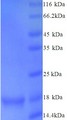
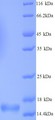
Greater than 92% by SDS-PAGE
Bio-Activity
Does not induce Lewy body inclusion formation in Sprague-Dawley rat primary hippocampal neurons. Thioflavin T emission curve shows only a small increase in fluorescence (indicative of alpha synuclein aggregation) when Type 2 alpha synuclein PFFs are combined with alpha synuclein monomers. Certain biological activities in other neuronal cells cannot be ruled out. Researchers should test compatibility prior to use.
Endotoxin
Not Tested
Presentation
PBS, pH 7.4
Storage
Store at -80°C.
Restrictions
For research use only. Intended for use by laboratory professionals.
Available for shipment within the USA only
About SNCA / Alpha-Synuclein
Publications (0)
Customer Reviews (0)
Images
Immunofluorescence
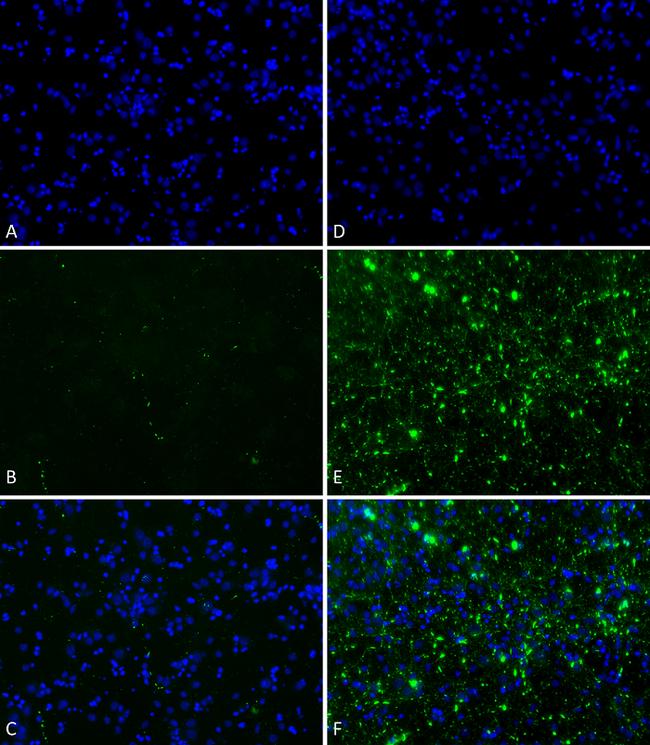
Primary rat hippocampal neurons show lewy body inclusion formation when treated with Type 1 Alpha Synuclein Preformed Fibrils at 4 µg/ml (D-F), but not when treated with Type 2 Alpha Synuclein Preformed Fibrils at 4 µg/ml (A-C). Tissue: Primary hippocampal neurons. Species: Sprague-Dawley rat. Fixation: 4% formaldehyde made from PFA. Primary Antibody: Mouse anti-pSer129 Antibody at 1:1000 24 hours at 4°C. Secondary Antibody: FITC Goat Anti-Mouse (green) at 1:700 for 1 hours at RT. Counterstain: Hoechst (blue) nuclear stain at 1:4000 for 1 hour at RT. Localization: Lewy body inclusions. Magnification: 20x.
Electron Microscopy

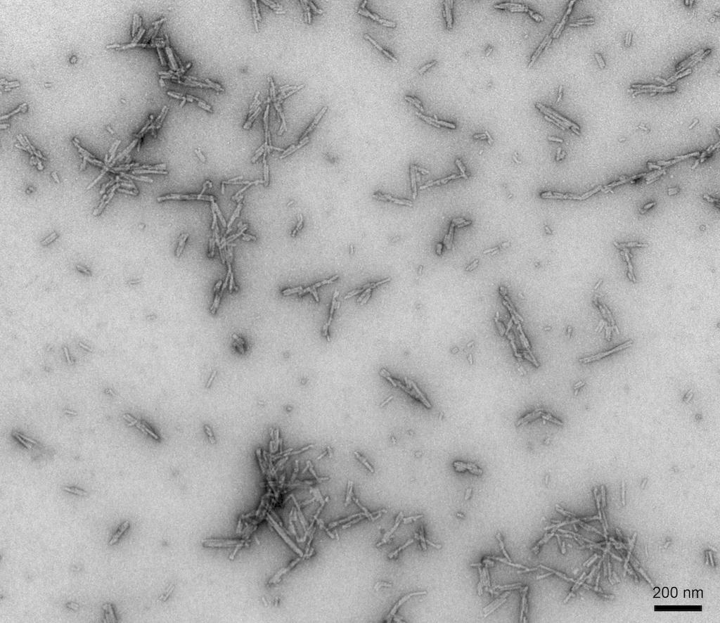
TEM of Type 2 Alpha Synuclein Preformed Fibrils (PFFs)
Functional Assay
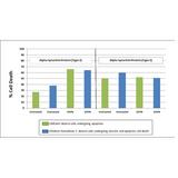
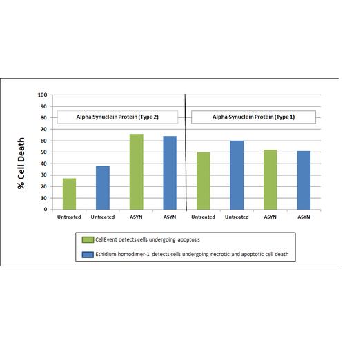
Toxicity results comparing Active Human Recombinant Alpha Synuclein Preformed Fibrils (Type 2) (Catalog No. SPR-317) and Active Human Recombinant Alpha Synuclein Preformed Fibrils (Type 1) (Catalog No. SPR-322). Data was graphed after live cell imaging results were obtained using the following procedure: After 8 days in vitro, primary rat mixed cortical neuron cells were washed with 1X PBS and treated with 500 µg/ml of Type 1 and Type 2 Alpha Synuclein Proteins for 20 hours at 37?C. Following treatements, cells were washed with 2X PBS and incubated with a staining solution (2.0 µM Cell Event + 2.5 µM Ethidium homodimer + 2.5 µg/ml Hoechst 33342 in sterile HBSS) for 30 minutes at 37?C. The addition of the Type 2 Alpha Synuclein Proteins resulted in a significant increase in cell death.
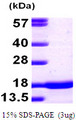
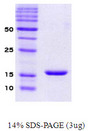
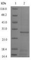
Sodium Dodecyl Sulfate - Polyacrylamide Gel Electrophoresis
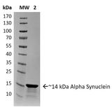
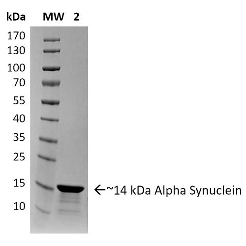
SDS-PAGE of ~14 kDa Human Recombinant Alpha Synuclein Protein Preformed Fibrils (Type 2). Lane 1: Molecular Weight Ladder (MW). Lane 2: Alpha Synuclein Protein Preformed Fibrils.
Immunofluorescence
Primary rat hippocampal neurons show lewy body inclusion formation when treated with Type 1 Alpha Synuclein Preformed Fibrils at 4 µg/ml (D-F), but not when treated with Type 2 Alpha Synuclein Preformed Fibrils at 4 µg/ml (A-C). Tissue: Primary hippocampal neurons. Species: Sprague-Dawley rat. Fixation: 4% formaldehyde made from PFA. Primary Antibody: Mouse anti-pSer129 Antibody at 1:1000 24 hours at 4°C. Secondary Antibody: FITC Goat Anti-Mouse (green) at 1:700 for 1 hours at RT. Counterstain: Hoechst (blue) nuclear stain at 1:4000 for 1 hour at RT. Localization: Lewy body inclusions. Magnification: 20x.
Electron Microscopy
TEM of Type 2 Alpha Synuclein Preformed Fibrils (PFFs)
Functional Assay
Toxicity results comparing Active Human Recombinant Alpha Synuclein Preformed Fibrils (Type 2) (Catalog No. SPR-317) and Active Human Recombinant Alpha Synuclein Preformed Fibrils (Type 1) (Catalog No. SPR-322). Data was graphed after live cell imaging results were obtained using the following procedure: After 8 days in vitro, primary rat mixed cortical neuron cells were washed with 1X PBS and treated with 500 µg/ml of Type 1 and Type 2 Alpha Synuclein Proteins for 20 hours at 37?C. Following treatements, cells were washed with 2X PBS and incubated with a staining solution (2.0 µM Cell Event + 2.5 µM Ethidium homodimer + 2.5 µg/ml Hoechst 33342 in sterile HBSS) for 30 minutes at 37?C. The addition of the Type 2 Alpha Synuclein Proteins resulted in a significant increase in cell death.
Sodium Dodecyl Sulfate - Polyacrylamide Gel Electrophoresis
SDS-PAGE of ~14 kDa Human Recombinant Alpha Synuclein Protein Preformed Fibrils (Type 2). Lane 1: Molecular Weight Ladder (MW). Lane 2: Alpha Synuclein Protein Preformed Fibrils.
Immunofluorescence
Primary rat hippocampal neurons show lewy body inclusion formation when treated with Type 1 Alpha Synuclein Preformed Fibrils at 4 µg/ml (D-F), but not when treated with Type 2 Alpha Synuclein Preformed Fibrils at 4 µg/ml (A-C). Tissue: Primary hippocampal neurons. Species: Sprague-Dawley rat. Fixation: 4% formaldehyde made from PFA. Primary Antibody: Mouse anti-pSer129 Antibody at 1:1000 24 hours at 4°C. Secondary Antibody: FITC Goat Anti-Mouse (green) at 1:700 for 1 hours at RT. Counterstain: Hoechst (blue) nuclear stain at 1:4000 for 1 hour at RT. Localization: Lewy body inclusions. Magnification: 20x.
Electron Microscopy
TEM of Type 2 Alpha Synuclein Preformed Fibrils (PFFs)
Functional Assay
Toxicity results comparing Active Human Recombinant Alpha Synuclein Preformed Fibrils (Type 2) (Catalog No. SPR-317) and Active Human Recombinant Alpha Synuclein Preformed Fibrils (Type 1) (Catalog No. SPR-322). Data was graphed after live cell imaging results were obtained using the following procedure: After 8 days in vitro, primary rat mixed cortical neuron cells were washed with 1X PBS and treated with 500 µg/ml of Type 1 and Type 2 Alpha Synuclein Proteins for 20 hours at 37?C. Following treatements, cells were washed with 2X PBS and incubated with a staining solution (2.0 µM Cell Event + 2.5 µM Ethidium homodimer + 2.5 µg/ml Hoechst 33342 in sterile HBSS) for 30 minutes at 37?C. The addition of the Type 2 Alpha Synuclein Proteins resulted in a significant increase in cell death.
Sodium Dodecyl Sulfate - Polyacrylamide Gel Electrophoresis
SDS-PAGE of ~14 kDa Human Recombinant Alpha Synuclein Protein Preformed Fibrils (Type 2). Lane 1: Molecular Weight Ladder (MW). Lane 2: Alpha Synuclein Protein Preformed Fibrils.
Popular SNCA / Alpha-Synuclein Proteins
Source:
Yeast
Tag:
6His, N-terminus
Source:
Yeast
Tag:
His
Source:
E. coli
Tag:
GST, N-terminus
Request SDS/MSDS
To request an SDS/MSDS form for this product, please contact our Technical Support department at:
Technical.Support@LSBio.com
Requested From: United States
Date Requested: 5/14/2024
Date Requested: 5/14/2024


