order histories, retained contact details for faster checkout, review submissions, and special promotions.
Forgot password?
order histories, retained contact details for faster checkout, review submissions, and special promotions.
Locations
Orders Processing,
Shipping & Receiving,
Warehouse
2 Shaker Rd Suites
B001/B101
Shirley, MA 01464
Production Lab
Floor 6, Suite 620
20700 44th Avenue W
Lynnwood, WA 98036
Telephone Numbers
Tel: +1 (206) 374-1102
Fax: +1 (206) 577-4565
Contact Us
Additional Contact Details
order histories, retained contact details for faster checkout, review submissions, and special promotions.
Forgot password?
order histories, retained contact details for faster checkout, review submissions, and special promotions.
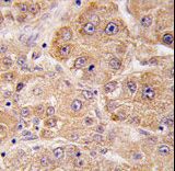
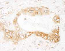
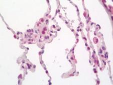
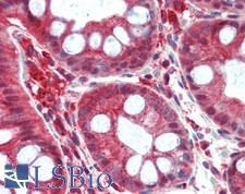
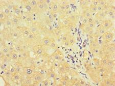
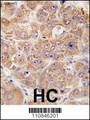
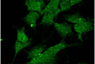
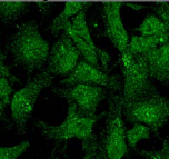
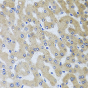
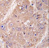
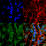
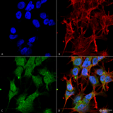
UVRAG
UV radiation resistance associated
UVRAG complements the ultraviolet sensitivity of xeroderma pigmentosum group C cells and encodes a protein with a C2 domain. The protein activates the Beclin1-PI(3)KC3 complex, promoting autophagy and suppressing the proliferation and tumorigenicity of human colon cancer cells. Chromosomal aberrations involving this gene are associated with left-right axis malformation and mutations in this gene have been associated with colon cancer.
| Gene Name: | UV radiation resistance associated |
| Synonyms: | UVRAG, DHTX, Beclin 1 binding protein, VPS38, Disrupted in heterotaxy, p63 |
| Target Sequences: | NM_003369 NP_003360.2 Q9P2Y5 |

![UVRAG Antibody - Immunofluorescence of monoclonal antibody to UVRAG on HeLa cell . [antibody concentration 10 ug/ml]](https://lsbio-7d62.kxcdn.com/image2/uvrag-antibody-clone-2e8-ls-c198419/146935_5044452.jpg)


If you do not find the reagent or information you require, please contact Customer.Support@LSBio.com to inquire about additional products in development.


