order histories, retained contact details for faster checkout, review submissions, and special promotions.
Forgot password?
order histories, retained contact details for faster checkout, review submissions, and special promotions.
Locations
Orders Processing,
Shipping & Receiving,
Warehouse
2 Shaker Rd Suites
B001/B101
Shirley, MA 01464
Production Lab
Floor 6, Suite 620
20700 44th Avenue W
Lynnwood, WA 98036
Telephone Numbers
Tel: +1 (206) 374-1102
Fax: +1 (206) 577-4565
Contact Us
Additional Contact Details
order histories, retained contact details for faster checkout, review submissions, and special promotions.
Forgot password?
order histories, retained contact details for faster checkout, review submissions, and special promotions.
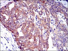
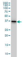
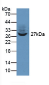
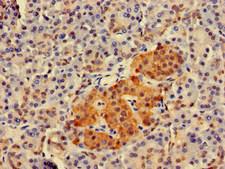
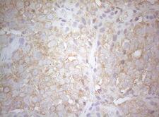
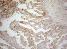
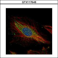
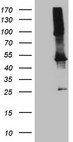
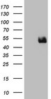
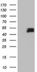
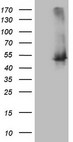
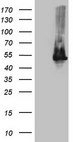
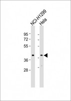
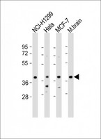
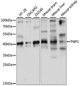
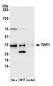
TWF1 / PTK9
twinfilin actin-binding protein 1
Actin-binding protein involved in motile and morphological processes. Inhibits actin polymerization, likely by sequestering G-actin. By capping the barbed ends of filaments, it also regulates motility. Seems to play an important role in clathrin-mediated endocytosis and distribution of endocytic organelles.
| Gene Name: | twinfilin actin-binding protein 1 |
| Family/Subfamily: | Protein Kinase , A6 |
| Synonyms: | TWF1, A6, A6 protein tyrosine kinase, Protein A6, Twinfilin, Twinfilin-1, Protein tyrosine kinase, Protein tyrosine kinase 9, PTK9, PTK9 protein tyrosine kinase 9 |
| Target Sequences: | NM_002822 NP_002813.3 Q12792 |


If you do not find the reagent or information you require, please contact Customer.Support@LSBio.com to inquire about additional products in development.


