order histories, retained contact details for faster checkout, review submissions, and special promotions.
Forgot password?
order histories, retained contact details for faster checkout, review submissions, and special promotions.
Locations
Orders Processing,
Shipping & Receiving,
Warehouse
2 Shaker Rd Suites
B001/B101
Shirley, MA 01464
Production Lab
Floor 6, Suite 620
20700 44th Avenue W
Lynnwood, WA 98036
Telephone Numbers
Tel: +1 (206) 374-1102
Fax: +1 (206) 577-4565
Contact Us
Additional Contact Details
order histories, retained contact details for faster checkout, review submissions, and special promotions.
Forgot password?
order histories, retained contact details for faster checkout, review submissions, and special promotions.
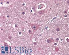
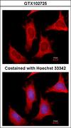
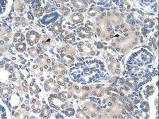
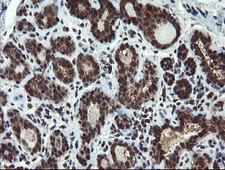
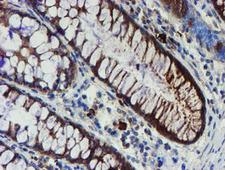
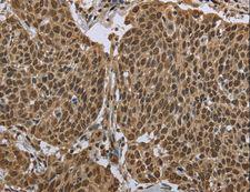
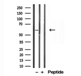
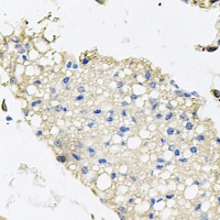
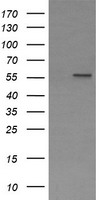
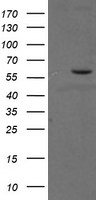
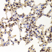
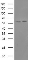
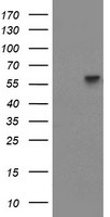
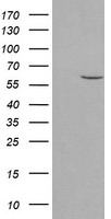
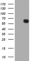
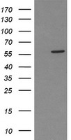
PPAT
phosphoribosyl pyrophosphate amidotransferase
The protein encoded by this gene is a member of the purine/pyrimidine phosphoribosyltransferase family. It is a regulatory allosteric enzyme that catalyzes the first step of de novo purine nucleotide biosythetic pathway. This gene and PAICS/AIRC gene, a bifunctional enzyme catalyzing steps six and seven of this pathway, are located in close proximity on chromosome 4, and divergently transcribed from an intergenic region.
| Gene Name: | phosphoribosyl pyrophosphate amidotransferase |
| Synonyms: | PPAT, Amidophosphoribosyltransferase, ATASE, GPAT, PRAT |
| Target Sequences: | NM_002703 NP_002694.3 Q06203 |
Publications (2)


If you do not find the reagent or information you require, please contact Customer.Support@LSBio.com to inquire about additional products in development.


