Login
Registration enables users to use special features of this website, such as past
order histories, retained contact details for faster checkout, review submissions, and special promotions.
order histories, retained contact details for faster checkout, review submissions, and special promotions.
Forgot password?
Registration enables users to use special features of this website, such as past
order histories, retained contact details for faster checkout, review submissions, and special promotions.
order histories, retained contact details for faster checkout, review submissions, and special promotions.
Quick Order
Products
Antibodies
ELISA and Assay Kits
Research Areas
Infectious Disease
Resources
Purchasing
Reference Material
Contact Us
Locations
Orders Processing,
Shipping & Receiving,
Warehouse
2 Shaker Rd Suites
B001/B101
Shirley, MA 01464
Production Lab
Floor 6, Suite 620
20700 44th Avenue W
Lynnwood, WA 98036
Telephone Numbers
Tel: +1 (206) 374-1102
Fax: +1 (206) 577-4565
Contact Us
Additional Contact Details
Login
Registration enables users to use special features of this website, such as past
order histories, retained contact details for faster checkout, review submissions, and special promotions.
order histories, retained contact details for faster checkout, review submissions, and special promotions.
Forgot password?
Registration enables users to use special features of this website, such as past
order histories, retained contact details for faster checkout, review submissions, and special promotions.
order histories, retained contact details for faster checkout, review submissions, and special promotions.
Quick Order
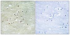
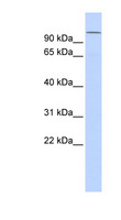
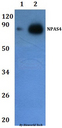
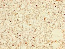
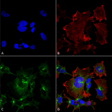
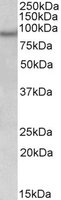
NXF / NPAS4
neuronal PAS domain protein 4
Acts as a transcriptional activator in the presence of ARNT. Can activate the CME (CNS midline enhancer) element and the expression of the drebrin gene.
| Gene Name: | neuronal PAS domain protein 4 |
| Synonyms: | NPAS4, BHLHe79, Neuronal PAS domain protein 4, Neuronal PAS4, Le-PAS, NXF, PASD10 |
| Target Sequences: | NP_849195.2 Q8IUM7 |
☰ Filters
Products
Antibodies
(23)
Type
Primary
(23)
Target
NXF / NPAS4
(23)
Reactivity
Human
(22)
Mouse
(17)
Rat
(15)
Bat
(1)
Bovine
(4)
Chimpanzee
(3)
Dog
(4)
Gibbon
(3)
Hamster
(1)
Horse
(4)
Monkey
(5)
Pig
(5)
Rabbit
(4)
Application
IHC
(13)
IHC-P
(2)
WB
(17)
ELISA
(3)
ICC
(1)
IF
(9)
Peptide-ELISA
(2)
Host
rabbit
(14)
mouse
(8)
goat
(1)
Isotype
IgG
(7)
IgG1
(8)
Clonality
monoclonal mc
(8)
polyclonal pc
(15)
Clone
S408-79
(8)
Format
APC Conjugated
(1)
Atto 390 Conjugated
(1)
Atto 594 Conjugated
(1)
Biotin Conjugated
(3)
FITC Conjugated
(3)
HRP Conjugated
(2)
PerCP Conjugated
(1)
RPE Conjugated
(1)
Unconjugated
(10)
Epitope
aa539-588
(4)
C-Terminus
(2)
580-660 aa, C-terminal
(1)
aa149-161
(1)
aa603-652
(1)
Publications
No
(23)

NXF / NPAS4 Rabbit anti-Human Polyclonal Antibody
Mouse, Rat, Human
ELISA, IHC
Unconjugated
100 µg/$357
NXF / NPAS4 Rabbit anti-Human Polyclonal (aa603-652) Antibody
Mouse, Rat, Human
IHC, Peptide-ELISA
Unconjugated
50 µl/$334; 100 µl/$397

NXF / NPAS4 Rabbit anti-Human Polyclonal (aa539-588) Antibody
Human
WB
Unconjugated
100 µl/$424

NXF / NPAS4 Rabbit anti-Human Polyclonal (aa539-588) (HRP) Antibody
Rabbit, Mouse, Dog, Bovine, Rat, Pig, Horse, Gibbon, Chimpanzee, Human, Monkey
WB
HRP Conjugated
100 µl/$469

NXF / NPAS4 Rabbit anti-Human Polyclonal (aa539-588) (Biotin) Antibody
Rabbit, Mouse, Dog, Bovine, Rat, Pig, Horse, Gibbon, Chimpanzee, Human, Monkey
WB
Biotin Conjugated
100 µl/$469
NXF / NPAS4 Rabbit anti-Human Polyclonal (aa539-588) (FITC) Antibody
Rabbit, Mouse, Dog, Bovine, Rat, Pig, Horse, Gibbon, Chimpanzee, Human, Monkey
WB
FITC Conjugated
100 µl/$469
NXF / NPAS4 Rabbit anti-Human Polyclonal Antibody
Mouse, Human
WB
Unconjugated
50 µl/$321; 100 µl/$403
NXF / NPAS4 Rabbit anti-Human Polyclonal Antibody
Human
ELISA, IHC, IHC-P, WB
Unconjugated
50 µg/$294; 100 µg/$360
NXF / NPAS4 Rabbit anti-Human Polyclonal (HRP) Antibody
Human
HRP Conjugated
50 µg/$294; 100 µg/$360
NXF / NPAS4 Rabbit anti-Human Polyclonal (Biotin) Antibody
Human
Biotin Conjugated
50 µg/$294; 100 µg/$360
NXF / NPAS4 Rabbit anti-Human Polyclonal (FITC) Antibody
Human
FITC Conjugated
50 µg/$294; 100 µg/$360

NXF / NPAS4 Mouse anti-Human Monoclonal (S408-79) Antibody
Mouse, Rat, Human
IF, IHC, WB
Unconjugated
100 µg/$442

NXF / NPAS4 Mouse anti-Human Monoclonal (Biotin) (S408-79) Antibody
Mouse, Rat, Human
IF, IHC, WB
Biotin Conjugated
100 µg/$475

NXF / NPAS4 Mouse anti-Human Monoclonal (FITC) (S408-79) Antibody
Mouse, Rat, Human
IF, IHC, WB
FITC Conjugated
100 µg/$472

NXF / NPAS4 Mouse anti-Human Monoclonal (APC) (S408-79) Antibody
Mouse, Rat, Human
IF, IHC, WB
APC Conjugated
100 µg/$478
NXF / NPAS4 Mouse anti-Human Monoclonal (RPE) (S408-79) Antibody
Mouse, Rat, Human
IF, IHC, WB
RPE Conjugated
100 µg/$477

NXF / NPAS4 Mouse anti-Human Monoclonal (Atto 390) (S408-79) Antibody
Mouse, Rat, Human
IF, IHC, WB
Atto 390 Conjugated
100 µg/$479

NXF / NPAS4 Mouse anti-Human Monoclonal (PerCP) (S408-79) Antibody
Mouse, Rat, Human
IF, IHC, WB
PerCP Conjugated
100 µg/$478

NXF / NPAS4 Mouse anti-Human Monoclonal (Atto 594) (S408-79) Antibody
Mouse, Rat, Human
IF, IHC, WB
Atto 594 Conjugated
100 µg/$479
NXF / NPAS4 Rabbit anti-Human Polyclonal (580-660 aa, C-terminal) Antibody
Mouse, Rat, Human
ELISA, IHC
Unconjugated
50 µg/$295; 100 µg/$335; 200 µg/$394
NXF / NPAS4 Rabbit anti-Mouse Polyclonal (C-Terminus) Antibody
Mouse
WB
Unconjugated
100 µl/$440; 200 µl/$468
NXF / NPAS4 Goat anti-Human Polyclonal (aa149-161) Antibody
Rabbit, Mouse, Dog, Bovine, Rat, Hamster, Pig, Horse, Bat, Human, Monkey
Peptide-ELISA, WB
Unconjugated
100 µg/$503
Viewing 26-23
of 23
product results
If you do not find the reagent or information you require, please contact Customer.Support@LSBio.com to inquire about additional products in development.


