order histories, retained contact details for faster checkout, review submissions, and special promotions.
Forgot password?
order histories, retained contact details for faster checkout, review submissions, and special promotions.
Locations
Orders Processing,
Shipping & Receiving,
Warehouse
2 Shaker Rd Suites
B001/B101
Shirley, MA 01464
Production Lab
Floor 6, Suite 620
20700 44th Avenue W
Lynnwood, WA 98036
Telephone Numbers
Tel: +1 (206) 374-1102
Fax: +1 (206) 577-4565
Contact Us
Additional Contact Details
order histories, retained contact details for faster checkout, review submissions, and special promotions.
Forgot password?
order histories, retained contact details for faster checkout, review submissions, and special promotions.
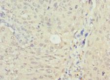
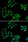
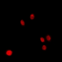
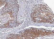
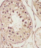
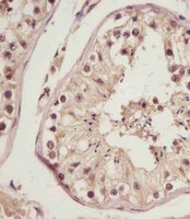
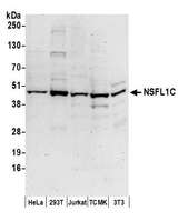
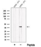
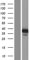
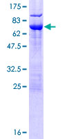
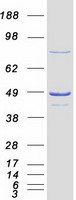
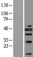
NSFL1C
NSFL1 (p97) Cofactor (p47)
N-ethylmaleimide-sensitive factor (NSF) and valosin-containing protein (p97) are two ATPases known to be involved in transport vesicle/target membrane fusion and fusions between membrane compartments. A trimer of the protein encoded by this gene binds a hexamer of cytosolic p97 and is required for p97-mediated regrowth of Golgi cisternae from mitotic Golgi fragments. Alternative splicing results in multiple transcript variants. A related pseudogene has been identified on chromosome 8.
| Gene Name: | NSFL1 (p97) Cofactor (p47) |
| Synonyms: | NSFL1C |
| Target Sequences: | NM_016143 NP_057227.2 Q9UNZ2 |

If you do not find the reagent or information you require, please contact Customer.Support@LSBio.com to inquire about additional products in development.


