order histories, retained contact details for faster checkout, review submissions, and special promotions.
Forgot password?
order histories, retained contact details for faster checkout, review submissions, and special promotions.
Locations
Orders Processing,
Shipping & Receiving,
Warehouse
2 Shaker Rd Suites
B001/B101
Shirley, MA 01464
Production Lab
Floor 6, Suite 620
20700 44th Avenue W
Lynnwood, WA 98036
Telephone Numbers
Tel: +1 (206) 374-1102
Fax: +1 (206) 577-4565
Contact Us
Additional Contact Details
order histories, retained contact details for faster checkout, review submissions, and special promotions.
Forgot password?
order histories, retained contact details for faster checkout, review submissions, and special promotions.
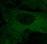
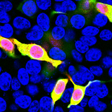
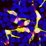
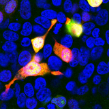
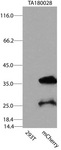
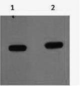
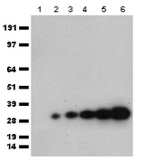
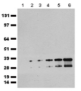
mCherry
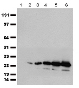
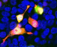
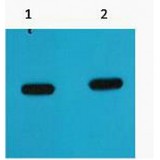
mCherry is a fluorophore used as a tracer to follow the flow of fluids, as a marker when tagged to molecules and cell components. mCherry and the majority of red fluorescent proteins derive from a protein isolated from Discosoma sp., while other fluorescent proteins in the green range are often variants of GFP from Aequorea victoria. mCherry is sometimes preferred to other fluorophores due to its color, as well as its photostability compared to other monomeric fluorophores. The 'm' in the name denotes its monomer configuration, which may be of importance in experimental design (other variants may be prefixed with 'td' for tandem-dimer, for example).
mCherry Target Details
| Target Name: | mCherry |
Publications (8)

If you do not find the reagent or information you require, please contact Customer.Support@LSBio.com to inquire about additional products in development.


