order histories, retained contact details for faster checkout, review submissions, and special promotions.
Forgot password?
order histories, retained contact details for faster checkout, review submissions, and special promotions.
Locations
Orders Processing,
Shipping & Receiving,
Warehouse
2 Shaker Rd Suites
B001/B101
Shirley, MA 01464
Production Lab
Floor 6, Suite 620
20700 44th Avenue W
Lynnwood, WA 98036
Telephone Numbers
Tel: +1 (206) 374-1102
Fax: +1 (206) 577-4565
Contact Us
Additional Contact Details
order histories, retained contact details for faster checkout, review submissions, and special promotions.
Forgot password?
order histories, retained contact details for faster checkout, review submissions, and special promotions.
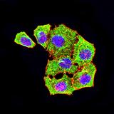
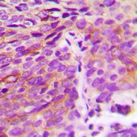
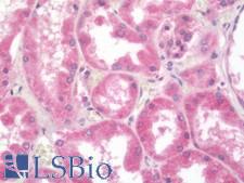
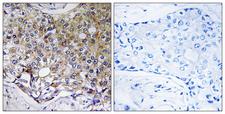
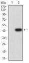
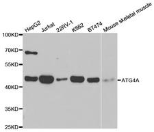
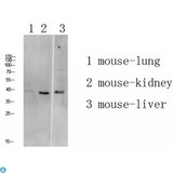
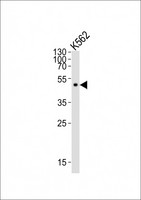
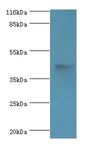
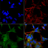
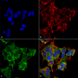
ATG4A
autophagy related 4A, cysteine peptidase
Cysteine protease required for the cytoplasm to vacuole transport (Cvt) and autophagy. Cleaves the C-terminal amino acid of ATG8 family proteins to reveal a C-terminal glycine. Exposure of the glycine at the C-terminus is essential for ATG8 proteins conjugation to phosphatidylethanolamine (PE) and insertion to membranes, which is necessary for autophagy. Preferred substrate is GABARAPL2 followed by MAP1LC3A and GABARAP. Has also an activity of delipidating enzyme for the PE-conjugated forms.
| Gene Name: | autophagy related 4A, cysteine peptidase |
| Synonyms: | ATG4A, AUTL2, Autophagin-2, APG4A, Autophagin 2, Cysteine protease ATG4A, HAPG4A, APG4 autophagy 4 homolog A |
| Target Sequences: | NM_052936 Q8WYN0 Q8WYN0 |


If you do not find the reagent or information you require, please contact Customer.Support@LSBio.com to inquire about additional products in development.


