order histories, retained contact details for faster checkout, review submissions, and special promotions.
Forgot password?
order histories, retained contact details for faster checkout, review submissions, and special promotions.
Locations
Orders Processing,
Shipping & Receiving,
Warehouse
2 Shaker Rd Suites
B001/B101
Shirley, MA 01464
Production Lab
Floor 6, Suite 620
20700 44th Avenue W
Lynnwood, WA 98036
Telephone Numbers
Tel: +1 (206) 374-1102
Fax: +1 (206) 577-4565
Contact Us
Additional Contact Details
order histories, retained contact details for faster checkout, review submissions, and special promotions.
Forgot password?
order histories, retained contact details for faster checkout, review submissions, and special promotions.
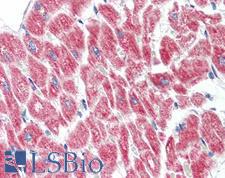
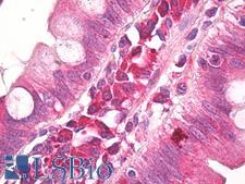
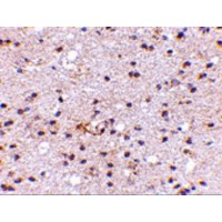
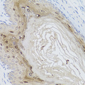
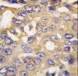
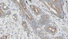
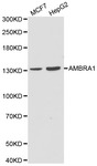
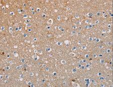
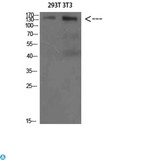
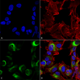
AMBRA1
autophagy/beclin-1 regulator 1
Regulates autophagy and development of the nervous system. Involved in autophagy in controlling protein turnover during neuronal development, and in regulating normal cell survival and proliferation.
| Gene Name: | autophagy/beclin-1 regulator 1 |
| Synonyms: | AMBRA1, Autophagy/beclin-1 regulator 1, KIAA1736, WD repeat domain 94, WDR94, DCAF3 |
| Target Sequences: | AK023197 BAB14457.1 Q9C0C7 |


If you do not find the reagent or information you require, please contact Customer.Support@LSBio.com to inquire about additional products in development.


