Login
Registration enables users to use special features of this website, such as past
order histories, retained contact details for faster checkout, review submissions, and special promotions.
order histories, retained contact details for faster checkout, review submissions, and special promotions.
Forgot password?
Registration enables users to use special features of this website, such as past
order histories, retained contact details for faster checkout, review submissions, and special promotions.
order histories, retained contact details for faster checkout, review submissions, and special promotions.
Quick Order
Products
Antibodies
ELISA and Assay Kits
Research Areas
Infectious Disease
Resources
Purchasing
Reference Material
Contact Us
Locations
Orders Processing,
Shipping & Receiving,
Warehouse
2 Shaker Rd Suites
B001/B101
Shirley, MA 01464
Production Lab
Floor 6, Suite 620
20700 44th Avenue W
Lynnwood, WA 98036
Telephone Numbers
Tel: +1 (206) 374-1102
Fax: +1 (206) 577-4565
Contact Us
Additional Contact Details
Login
Registration enables users to use special features of this website, such as past
order histories, retained contact details for faster checkout, review submissions, and special promotions.
order histories, retained contact details for faster checkout, review submissions, and special promotions.
Forgot password?
Registration enables users to use special features of this website, such as past
order histories, retained contact details for faster checkout, review submissions, and special promotions.
order histories, retained contact details for faster checkout, review submissions, and special promotions.
Quick Order
| Catalog Number | Size | Price |
|---|---|---|
| LS-G3822-10 | 10 µg (0.2 mg/ml) | $351 |
| LS-G3822-50 | 50 µg | $564 |
1 of 2
2 of 2
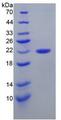
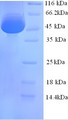
Human CALR / Calreticulin Protein (Recombinant His) (aa18-417) - LS-G3822
Human CALR / Calreticulin Protein (Recombinant His) (aa18-417) - LS-G3822
Description:
CALR / Calreticulin Protein LS-G3822 is a Recombinant Human CALR / Calreticulin produced in E. coli aa 18-417 with His tag(s). It is low in endotoxin; Less than 1.0 EU/µg protein (determined by LAL method). For Research Use Only
Toll Free North America
 206-374-1102
For Research Use Only
Overview
Description:
CALR / Calreticulin Protein LS-G3822 is a Recombinant Human CALR / Calreticulin produced in E. coli aa 18-417 with His tag(s). It is low in endotoxin; Less than 1.0 EU/µg protein (determined by LAL method). For Research Use Only
Specifications
Type
Recombinant Protein
Target
CALR / Calreticulin
Synonyms
CALR | Calregulin | CC1qR | CRP55 | CRT | CRTC | ERp60 | Grp60 | RO | SSA | Calreticulin | HACBP
Species
Human
Modifications
Unmodified
Conjugations
Unconjugated
Tag
His
Region
aa 18-417
Predicted Molecular Weight
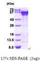
~55kDa (SDS-PAGE)
Expression System
E. coli
Source Species
E. coli
Purification
Greater than 90% by SDS-PAGE
Bio-Activity
Not Tested
Endotoxin
Less than 1.0 EU/µg protein (determined by LAL method).
Presentation
55 mM Tris-HCl, pH 8.2, 150 mM NaCl
Storage
Store at 4°C for immediate use, or aliquot and store at -20°C for up to 3 months. Avoid freeze-thaw cycles.
Restrictions
For research use only. Intended for use by laboratory professionals.
About CALR / Calreticulin
Publications (0)
Customer Reviews (0)
Images
Functional Assay
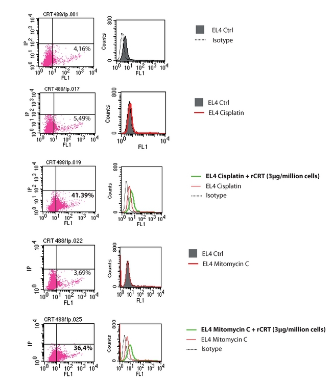
Flow cytometric analysis of CRT on the cell surface 3.10^5 EL4 Thymoma cells, growing in suspension in RPMI 1640 (Gibco) supplemented medium were plated in 12-well plates and treated with mitomycin C (30mM, Sanofi Aventis) or cisplatin (25mM, Sigma) for 4h. Cells were harvested, washed once with cold PBS and possibly resuspended in 200mL of cold PBS containing 1mg of recombinant Calreticulin for 30 minutesutes on ice. After one wash with cold PBS, cells were fixed in 0.25% paraformaldehyde (PFA) in PBS for 5 minutesutes. After washing again once with cold PBS, cells were incubated for 30 minutes with primary antibody, diluted in cold blocking buffer (2% FBS in PBS), followed by washing and incubation with the Alexa488-conjugated monoclonal secondary antibody in blocking buffer (30 minutes). Each sample was then analyzed by FACScan (Becton Dickinson) to identify cell-surface Calreticulin. Secondary antibody alone was used as an isotype control, and the fluorescent intensity of stained cells was gated on propridium iodide (PI) negative cells.Pictures courtesy of Prof. Guido Kroemer, INSERM, Paris.
Functional Assay

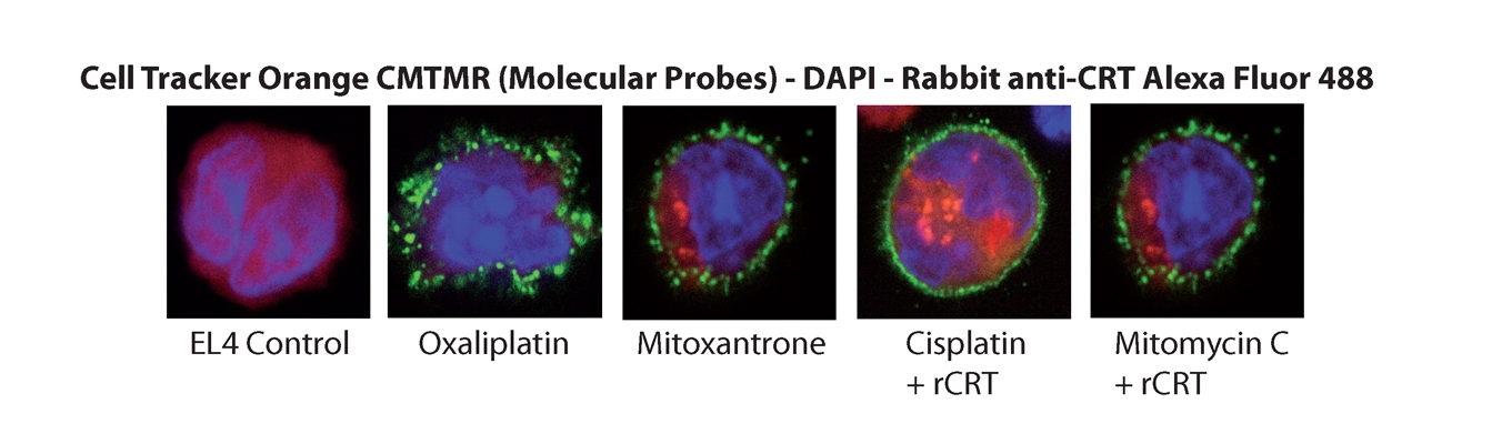
Immunofluorescence Cells were possibly incubated with rCRT and mitoxanthron (1mM, Sigma) treated cells were used as positive control. Pictures courtesy of Prof. Guido Kroemer, INSERM, Paris.
Functional Assay
Flow cytometric analysis of CRT on the cell surface 3.10^5 EL4 Thymoma cells, growing in suspension in RPMI 1640 (Gibco) supplemented medium were plated in 12-well plates and treated with mitomycin C (30mM, Sanofi Aventis) or cisplatin (25mM, Sigma) for 4h. Cells were harvested, washed once with cold PBS and possibly resuspended in 200mL of cold PBS containing 1mg of recombinant Calreticulin for 30 minutesutes on ice. After one wash with cold PBS, cells were fixed in 0.25% paraformaldehyde (PFA) in PBS for 5 minutesutes. After washing again once with cold PBS, cells were incubated for 30 minutes with primary antibody, diluted in cold blocking buffer (2% FBS in PBS), followed by washing and incubation with the Alexa488-conjugated monoclonal secondary antibody in blocking buffer (30 minutes). Each sample was then analyzed by FACScan (Becton Dickinson) to identify cell-surface Calreticulin. Secondary antibody alone was used as an isotype control, and the fluorescent intensity of stained cells was gated on propridium iodide (PI) negative cells.Pictures courtesy of Prof. Guido Kroemer, INSERM, Paris.
Functional Assay
Immunofluorescence Cells were possibly incubated with rCRT and mitoxanthron (1mM, Sigma) treated cells were used as positive control. Pictures courtesy of Prof. Guido Kroemer, INSERM, Paris.
Functional Assay
Flow cytometric analysis of CRT on the cell surface 3.10^5 EL4 Thymoma cells, growing in suspension in RPMI 1640 (Gibco) supplemented medium were plated in 12-well plates and treated with mitomycin C (30mM, Sanofi Aventis) or cisplatin (25mM, Sigma) for 4h. Cells were harvested, washed once with cold PBS and possibly resuspended in 200mL of cold PBS containing 1mg of recombinant Calreticulin for 30 minutesutes on ice. After one wash with cold PBS, cells were fixed in 0.25% paraformaldehyde (PFA) in PBS for 5 minutesutes. After washing again once with cold PBS, cells were incubated for 30 minutes with primary antibody, diluted in cold blocking buffer (2% FBS in PBS), followed by washing and incubation with the Alexa488-conjugated monoclonal secondary antibody in blocking buffer (30 minutes). Each sample was then analyzed by FACScan (Becton Dickinson) to identify cell-surface Calreticulin. Secondary antibody alone was used as an isotype control, and the fluorescent intensity of stained cells was gated on propridium iodide (PI) negative cells.Pictures courtesy of Prof. Guido Kroemer, INSERM, Paris.
Functional Assay
Immunofluorescence Cells were possibly incubated with rCRT and mitoxanthron (1mM, Sigma) treated cells were used as positive control. Pictures courtesy of Prof. Guido Kroemer, INSERM, Paris.
Popular CALR / Calreticulin Proteins
Source:
E. coli
Tag:
His
Source:
Yeast
Tag:
6His, N-terminus
Source:
Human
Tag:
Myc-DDK (Flag)
Request SDS/MSDS
To request an SDS/MSDS form for this product, please contact our Technical Support department at:
Technical.Support@LSBio.com
Requested From: United States
Date Requested: 4/25/2024
Date Requested: 4/25/2024


