Login
Registration enables users to use special features of this website, such as past
order histories, retained contact details for faster checkout, review submissions, and special promotions.
order histories, retained contact details for faster checkout, review submissions, and special promotions.
Forgot password?
Registration enables users to use special features of this website, such as past
order histories, retained contact details for faster checkout, review submissions, and special promotions.
order histories, retained contact details for faster checkout, review submissions, and special promotions.
Quick Order
Products
Antibodies
ELISA and Assay Kits
Research Areas
Infectious Disease
Resources
Purchasing
Reference Material
Contact Us
Locations
Orders Processing,
Shipping & Receiving,
Warehouse
2 Shaker Rd Suites
B001/B101
Shirley, MA 01464
Production Lab
Floor 6, Suite 620
20700 44th Avenue W
Lynnwood, WA 98036
Telephone Numbers
Tel: +1 (206) 374-1102
Fax: +1 (206) 577-4565
Contact Us
Additional Contact Details
Login
Registration enables users to use special features of this website, such as past
order histories, retained contact details for faster checkout, review submissions, and special promotions.
order histories, retained contact details for faster checkout, review submissions, and special promotions.
Forgot password?
Registration enables users to use special features of this website, such as past
order histories, retained contact details for faster checkout, review submissions, and special promotions.
order histories, retained contact details for faster checkout, review submissions, and special promotions.
Quick Order
| Catalog Number | Size | Price |
|---|---|---|
| LS-B10590-50 | 50 µg (1 mg/ml) | $515 |
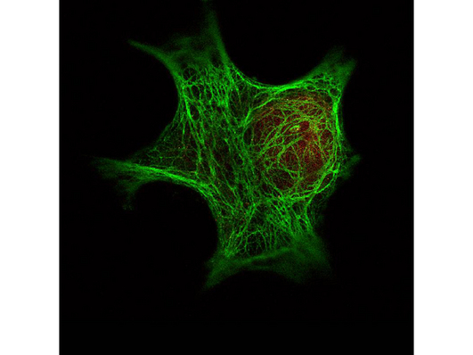
![Pan Cytokeratin Antibody - Immunofluorescence Microscopy - Anti-Keratin monoclonal antibody. Immunofluorescence using Mouse Anti-Keratin antibody. Confocal slices of HeLa cells are between 0.5 and 0.6 micron where the image is taken near the bottom of the cell. Use FITC a 1:2000 dilution of FITC conjugated Goat-a-Mouse IgG [H&L] (LS-C60676) for detection.](https://lsbio-7d62.kxcdn.com/image2/pathplus-pan-cytokeratin-antibody-clone-c11-ls-b10590/62644_90313.jpg)
![Pan Cytokeratin Antibody - Western blot - Anti-Keratin Monoclonal Antibody. Western blot of Mouse Anti-Keratin antibody. This antibody recognizes a single 56 kD band corresponding to human keratin as confirmed by the position of molecular weight markers (not shown). Approximatley 100 ng of keratin from human epidermis (Sigma K0253) was applied under reducing conditions to a pre-cast 4-20% iGel from Gradipore Inc. A 1:400 dilution of Mab anti-Keratin was used for 2h followed by detection using a 1:5000 dilution of IRDyeTM800 conjugated Goat-a-Mouse IgG [H&L] ( and visualization using the Odyssey Infrared Imaging System developed by LI-COR. Other detection systems will yield similar results. IRDye is a trademark of LI-COR, Inc.](https://lsbio-7d62.kxcdn.com/image2/pathplus-pan-cytokeratin-antibody-clone-c11-ls-b10590/62643_90312.jpg)
1 of 4
2 of 4
3 of 4
4 of 4
PathPlus™ Monoclonal Mouse anti‑Human Pan Cytokeratin Antibody (clone C11, IHC, IF, WB) LS‑B10590
PathPlus™ Monoclonal Mouse anti‑Human Pan Cytokeratin Antibody (clone C11, IHC, IF, WB) LS‑B10590

Note: This antibody replaces LS-C59516
Antibody:
Pan Cytokeratin Mouse anti-Human Monoclonal (C11) Antibody
Application:
IHC-P, IF, WB, IP, ELISA
Reactivity:
Human
Format:
Unconjugated, Unmodified
Toll Free North America
 206-374-1102
For Research Use Only
Overview
Antibody:
Pan Cytokeratin Mouse anti-Human Monoclonal (C11) Antibody
Application:
IHC-P, IF, WB, IP, ELISA
Reactivity:
Human
Format:
Unconjugated, Unmodified
Specifications
Host
Mouse
Reactivity
Human
(tested or 100% immunogen sequence identity)
Clonality
IgG1
Monoclonal
Clone
C11
Conjugations
Unconjugated
Purification
Protein A purified
Modifications
Unmodified
Immunogen
This protein A purified monoclonal antibody was produced by repeated immunizations with purified human cytoskeletal preparations from A431 cells.
Specificity
This antibody reacts with keratin (56 kDa), keratin 17 (46 kDa), keratin 18 (45 kDa) and keratin 19 (40 kDa) derived from humans. Cross reactivity with keratins from other sources has not been determined. No reaction is expected against other filament proteins including vimentin, desmin and neurofilament protein.
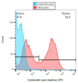
Applications
- IHC - Paraffin (10 µg/ml)
- Immunofluorescence (1:50 - 1:200)
- Western blot (1:50 - 1:200)
- Immunoprecipitation (1:100)
- ELISA (1:5000 - 1:20000)

|
Performing IHC? See our complete line of Immunohistochemistry Reagents including antigen retrieval solutions, blocking agents
ABC Detection Kits and polymers, biotinylated secondary antibodies, substrates and more.
|
Usage
This antibody is suitable for ELISA, immunohistochemistry, immunoblotting and immunoprecipitation. For a positive control use skin, colon carcinoma and squamous granulocyte carcinoma cells.
Presentation
0.02 M Potassium Phosphate, pH 7.2, 0.15 M NaCl, 0.01% Sodium Azide
Storage
Short term: store at 4°C. Long term: store at -20°C. Avoid freeze-thaw cycles.
Restrictions
For research use only. Intended for use by laboratory professionals.
Validation
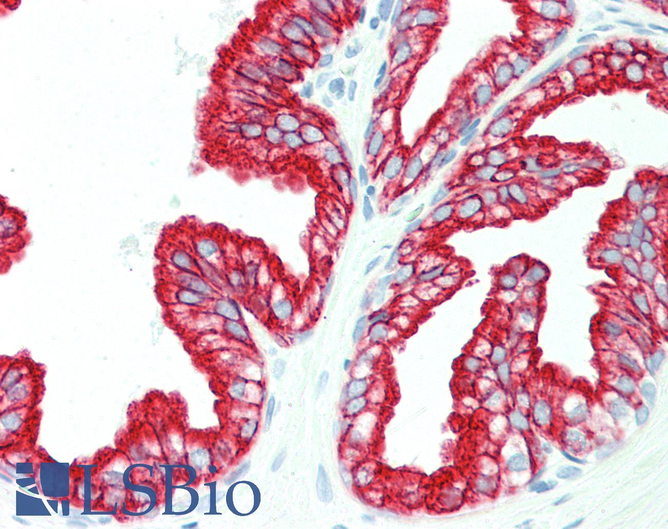
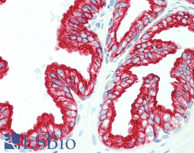
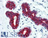
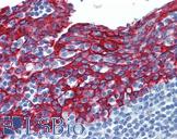
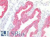
Anti-Pan Cytokeratin antibody IHC staining of human prostate. Immunohistochemistry of formalin-fixed, paraffin-embedded tissue after heat-induced antigen retrieval. Antibody concentration 10 ug/ml.
Anti-Pan Cytokeratin antibody IHC staining of human prostate. Immunohistochemistry of formalin-fixed, paraffin-embedded tissue after heat-induced antigen retrieval. Antibody concentration 10 ug/ml.
See More About...
LSBio Ratings
PathPlus™ Pan Cytokeratin Antibody (clone C11) for IHC, IF/Immunofluorescence, WB/Western, IP, ELISA LS-B10590 has an LSBio Rating of
Laboratory Validation Score (5)
Learn more about The LSBio Ratings Algorithm
Publications (0)
Customer Reviews (0)
Featured Products
Species:
Human
Applications:
IHC - Paraffin, Immunofluorescence, Western blot, ELISA
Species:
Human, Mammal
Applications:
IHC - Paraffin, ICC, Western blot, Immunoprecipitation, Flow Cytometry
Species:
Human
Applications:
IHC - Paraffin, Immunofluorescence, ELISA
Species:
Human, Mouse
Applications:
IHC - Paraffin, IHC - Frozen, ICC, Western blot
Species:
Human, Mammal
Applications:
IHC - Paraffin, ICC, Western blot, Immunoprecipitation, Flow Cytometry
Species:
Mammal
Applications:
IHC - Paraffin, ICC, Western blot, Immunoprecipitation, Flow Cytometry
Request SDS/MSDS
To request an SDS/MSDS form for this product, please contact our Technical Support department at:
Technical.Support@LSBio.com
Requested From: United States
Date Requested: 4/18/2024
Date Requested: 4/18/2024


