Login
Registration enables users to use special features of this website, such as past
order histories, retained contact details for faster checkout, review submissions, and special promotions.
order histories, retained contact details for faster checkout, review submissions, and special promotions.
Forgot password?
Registration enables users to use special features of this website, such as past
order histories, retained contact details for faster checkout, review submissions, and special promotions.
order histories, retained contact details for faster checkout, review submissions, and special promotions.
Quick Order
Products
Antibodies
ELISA and Assay Kits
Research Areas
Infectious Disease
Resources
Purchasing
Reference Material
Contact Us
Locations
Orders Processing,
Shipping & Receiving,
Warehouse
2 Shaker Rd Suites
B001/B101
Shirley, MA 01464
Production Lab
Floor 6, Suite 620
20700 44th Avenue W
Lynnwood, WA 98036
Telephone Numbers
Tel: +1 (206) 374-1102
Fax: +1 (206) 577-4565
Contact Us
Additional Contact Details
Login
Registration enables users to use special features of this website, such as past
order histories, retained contact details for faster checkout, review submissions, and special promotions.
order histories, retained contact details for faster checkout, review submissions, and special promotions.
Forgot password?
Registration enables users to use special features of this website, such as past
order histories, retained contact details for faster checkout, review submissions, and special promotions.
order histories, retained contact details for faster checkout, review submissions, and special promotions.
Quick Order
| Catalog Number | Size | Price |
|---|---|---|
| LS-K1071-30 | 30 Tests | $263 |
| LS-K1071-60 | 60 Tests | $502 |
![Apoptosis Assay Kit - DNA Fragmentation Detection Kit (LS-K1071) using paraffin fixed humantonsil tissue, 10 µm sections (1000X). [A] Section processed and counter-stainedwith methyl green according to the DNA Fragmentation Detection Kit manual. [B]Counter-stain step was eliminated to more clearly illustrate the level of positivestaining in the germinal centers of tonsil tissue. [C] Section treated with DNase I inorder to generate a positive control slide. Note all nuclei stain positive. The use ofDNase I generates free 3’-OH groups on cellular DNA, these free 3’-OH groupsare then labeled with biotin-nucleotide by the TdT in the DNA FragmentationDetection Kit (LS-K1071). [D] Negative control, where the TdT enzyme step waseliminated, thereby generating a negative slide.](https://lsbio-7d62.kxcdn.com/image2/dna-fragmentation-detection-kit-ls-k1071/515272_4325728.jpg)
1 of 2
2 of 2
DNA Fragmentation Detection Kit - LS-K1071
DNA Fragmentation Detection Kit - LS-K1071
Available for shipment within the USA only
Description:
Cell death occurs by two major mechanisms, necrosis and apoptosis. Apoptosis is also known as programmed cell death or anoikis (a form of apoptosis which is induced by anchorage-dependent cells detaching from the surrounding extracellular matrix). Apoptosis leads to the elimination of cells without releasing harmful substances into the surrounding area. Too little or too much apoptosis plays a role in a great many diseases. When apoptosis functions inappropriately, cells that should be eliminated survive and potentially become immortal, as in cancer or leukemia. When apoptosis works overly well, too many cells may ‘die’ and the result may be grave tissue damage. This is the case in stroke and neurodegenerative disorders such as Alzheimer, Huntington and Parkinson diseases. The term ‘apoptosis’ refers only to the structural changes a cell goes through during the process of programmed cell death and not to the process itself. Classical necrotic cell death occurs due to noxious injury or trauma to the cell while apoptosis is an energy dependent mechanism that takes place during normal cell development. While necrotic cell death results in cell lysis, cellular apoptosis is characterized morphologically by cell shrinkage, nuclear pyknosis, chromatin condensation, and blebbing of the plasma membrane. Apoptosis is the result of a cascade of molecular and biochemical events involving endogenous endonucleases that cleave DNA into the prototypical ‘ladder of DNA fragments’ that may be visualized in agarose gels. Observation of oligonucleosomal DNA fragments by DNA laddering has long been the most acceptable and only available assay for the detection of apoptosis. LSBio's DNA Fragmentation Detection Kit exploits the fact that apoptotic endonucleases not only affect cellular DNA by producing the classical DNA ladder but also generate free 3’-OH groups at the ends of these DNA fragments. These free 3’-OH groups are end-labeled by the DNA Fragmentation Detection Kit allowing for the detection of apoptotic cells using a molecular biology-based, end-labeling technique.
Required Components Not Supplied
- DNase I (not available through LSBio)
Available for USA Shipment Only
Toll Free North America
 206-374-1102
For Research Use Only
Overview
Description:
Cell death occurs by two major mechanisms, necrosis and apoptosis. Apoptosis is also known as programmed cell death or anoikis (a form of apoptosis which is induced by anchorage-dependent cells detaching from the surrounding extracellular matrix). Apoptosis leads to the elimination of cells without releasing harmful substances into the surrounding area. Too little or too much apoptosis plays a role in a great many diseases. When apoptosis functions inappropriately, cells that should be eliminated survive and potentially become immortal, as in cancer or leukemia. When apoptosis works overly well, too many cells may ‘die’ and the result may be grave tissue damage. This is the case in stroke and neurodegenerative disorders such as Alzheimer, Huntington and Parkinson diseases. The term ‘apoptosis’ refers only to the structural changes a cell goes through during the process of programmed cell death and not to the process itself. Classical necrotic cell death occurs due to noxious injury or trauma to the cell while apoptosis is an energy dependent mechanism that takes place during normal cell development. While necrotic cell death results in cell lysis, cellular apoptosis is characterized morphologically by cell shrinkage, nuclear pyknosis, chromatin condensation, and blebbing of the plasma membrane. Apoptosis is the result of a cascade of molecular and biochemical events involving endogenous endonucleases that cleave DNA into the prototypical ‘ladder of DNA fragments’ that may be visualized in agarose gels. Observation of oligonucleosomal DNA fragments by DNA laddering has long been the most acceptable and only available assay for the detection of apoptosis. LSBio's DNA Fragmentation Detection Kit exploits the fact that apoptotic endonucleases not only affect cellular DNA by producing the classical DNA ladder but also generate free 3’-OH groups at the ends of these DNA fragments. These free 3’-OH groups are end-labeled by the DNA Fragmentation Detection Kit allowing for the detection of apoptotic cells using a molecular biology-based, end-labeling technique.
Specifications
Name
DNA Fragmentation Detection Kit
Type
Detection/Quantition
Target
Apoptosis
SampleType
Cell Cultures, Tissues
Supplied Components
The following components are supplied with this product.
- Proteinase K
- TdT Equilibration Buffer
- TdT Labeling Reaction Mix
- TdT Enzyme
- Stop Buffer
- Block Buffer
- Streptavidin-HRP Conjugate (25X)
- DAB Concentrate
- DAB Reaction Buffer
- Methyl Green Counterstain
- (See Datasheet for specific volumes supplied)
Required Materials Not Supplied
The following materials are also required in order to run this assay.
- DNase I (not available through LSBio)
Applications
Microscopy (visible)
Equipment
Microscope (visible)
Conditions
Shipped +4°C Ice Packs, Store at -20°C
Documents
Restrictions
For research use only. Intended for use by laboratory professionals.
Available for shipment within the USA only
Guarantee
This Assay Kit carries the LSBio 100% Guarantee
Publications (0)
Customer Reviews (0)
Images
Microscopy (visible)

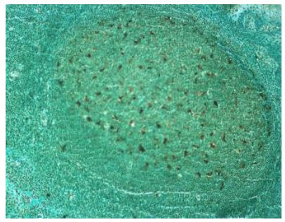
Positive result in DNA Fragmentation Detection Kit (LS-K1071) using paraffin fixed human tonsil tissue, 10 µm sections (1000X).
Microscopy (visible)
![Apoptosis Assay Kit - DNA Fragmentation Detection Kit (LS-K1071) using paraffin fixed humantonsil tissue, 10 µm sections (1000X). [A] Section processed and counter-stainedwith methyl green according to the DNA Fragmentation Detection Kit manual. [B]Counter-stain step was eliminated to more clearly illustrate the level of positivestaining in the germinal centers of tonsil tissue. [C] Section treated with DNase I inorder to generate a positive control slide. Note all nuclei stain positive. The use ofDNase I generates free 3’-OH groups on cellular DNA, these free 3’-OH groupsare then labeled with biotin-nucleotide by the TdT in the DNA FragmentationDetection Kit (LS-K1071). [D] Negative control, where the TdT enzyme step waseliminated, thereby generating a negative slide.](https://lsbio-7d62.kxcdn.com/image2/dna-fragmentation-detection-kit-ls-k1071/515272_4325727.jpg)
DNA Fragmentation Detection Kit (LS-K1071) using paraffin fixed humantonsil tissue, 10 µm sections (1000X). [A] Section processed and counter-stainedwith methyl green according to the DNA Fragmentation Detection Kit manual. [B]Counter-stain step was eliminated to more clearly illustrate the level of positivestaining in the germinal centers of tonsil tissue. [C] Section treated with DNase I inorder to generate a positive control slide. Note all nuclei stain positive. The use ofDNase I generates free 3’-OH groups on cellular DNA, these free 3’-OH groupsare then labeled with biotin-nucleotide by the TdT in the DNA FragmentationDetection Kit (LS-K1071). [D] Negative control, where the TdT enzyme step waseliminated, thereby generating a negative slide.
Microscopy (visible)
Positive result in DNA Fragmentation Detection Kit (LS-K1071) using paraffin fixed human tonsil tissue, 10 µm sections (1000X).
Microscopy (visible)
![Apoptosis Assay Kit - DNA Fragmentation Detection Kit (LS-K1071) using paraffin fixed humantonsil tissue, 10 µm sections (1000X). [A] Section processed and counter-stainedwith methyl green according to the DNA Fragmentation Detection Kit manual. [B]Counter-stain step was eliminated to more clearly illustrate the level of positivestaining in the germinal centers of tonsil tissue. [C] Section treated with DNase I inorder to generate a positive control slide. Note all nuclei stain positive. The use ofDNase I generates free 3’-OH groups on cellular DNA, these free 3’-OH groupsare then labeled with biotin-nucleotide by the TdT in the DNA FragmentationDetection Kit (LS-K1071). [D] Negative control, where the TdT enzyme step waseliminated, thereby generating a negative slide.](https://lsbio-7d62.kxcdn.com/image2/dna-fragmentation-detection-kit-ls-k1071/515272_4276627.jpg)
DNA Fragmentation Detection Kit (LS-K1071) using paraffin fixed humantonsil tissue, 10 µm sections (1000X). [A] Section processed and counter-stainedwith methyl green according to the DNA Fragmentation Detection Kit manual. [B]Counter-stain step was eliminated to more clearly illustrate the level of positivestaining in the germinal centers of tonsil tissue. [C] Section treated with DNase I inorder to generate a positive control slide. Note all nuclei stain positive. The use ofDNase I generates free 3’-OH groups on cellular DNA, these free 3’-OH groupsare then labeled with biotin-nucleotide by the TdT in the DNA FragmentationDetection Kit (LS-K1071). [D] Negative control, where the TdT enzyme step waseliminated, thereby generating a negative slide.
Microscopy (visible)
Positive result in DNA Fragmentation Detection Kit (LS-K1071) using paraffin fixed human tonsil tissue, 10 µm sections (1000X).
Microscopy (visible)
DNA Fragmentation Detection Kit (LS-K1071) using paraffin fixed humantonsil tissue, 10 µm sections (1000X). [A] Section processed and counter-stainedwith methyl green according to the DNA Fragmentation Detection Kit manual. [B]Counter-stain step was eliminated to more clearly illustrate the level of positivestaining in the germinal centers of tonsil tissue. [C] Section treated with DNase I inorder to generate a positive control slide. Note all nuclei stain positive. The use ofDNase I generates free 3’-OH groups on cellular DNA, these free 3’-OH groupsare then labeled with biotin-nucleotide by the TdT in the DNA FragmentationDetection Kit (LS-K1071). [D] Negative control, where the TdT enzyme step waseliminated, thereby generating a negative slide.
Request SDS/MSDS
To request an SDS/MSDS form for this product, please contact our Technical Support department at:
Technical.Support@LSBio.com
Requested From: United States
Date Requested: 4/25/2024
Date Requested: 4/25/2024


