Login
Registration enables users to use special features of this website, such as past
order histories, retained contact details for faster checkout, review submissions, and special promotions.
order histories, retained contact details for faster checkout, review submissions, and special promotions.
Forgot password?
Registration enables users to use special features of this website, such as past
order histories, retained contact details for faster checkout, review submissions, and special promotions.
order histories, retained contact details for faster checkout, review submissions, and special promotions.
Quick Order
Products
Antibodies
ELISA and Assay Kits
Research Areas
Infectious Disease
Resources
Purchasing
Reference Material
Contact Us
Locations
Orders Processing,
Shipping & Receiving,
Warehouse
2 Shaker Rd Suites
B001/B101
Shirley, MA 01464
Production Lab
Floor 6, Suite 620
20700 44th Avenue W
Lynnwood, WA 98036
Telephone Numbers
Tel: +1 (206) 374-1102
Fax: +1 (206) 577-4565
Contact Us
Additional Contact Details
Login
Registration enables users to use special features of this website, such as past
order histories, retained contact details for faster checkout, review submissions, and special promotions.
order histories, retained contact details for faster checkout, review submissions, and special promotions.
Forgot password?
Registration enables users to use special features of this website, such as past
order histories, retained contact details for faster checkout, review submissions, and special promotions.
order histories, retained contact details for faster checkout, review submissions, and special promotions.
Quick Order
| Catalog Number | Size | Price |
|---|---|---|
| LS-C18912-500 | 500 µg | $606 |
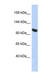
![UBA1 / UBE1 Antibody - Anti-Ubiquitin Activating Enzyme (E1) Antibody - Western Blot. Western blot of purified anti-Ubiquitin Activating Enzyme (E1) antibody shows detection of a band at ~118 kD corres-ponding to UBE1 (lane 1 800 nm channel). Approximately 35 ug of an A431 whole cell lysate was separated on a 4-20% Tris-Glycine gel by SDS-PAGE and transferred onto nitrocellulose. After blocking the membrane was probed with the primary antibody diluted to 1:1000. Incubation was for 2 h at room temperature followed by washes and reaction with a 1:10000 dilution of IRDye800 conjugated Gt-a-Rabbit IgG [H&L] MX10 ( for 45 min at room temperature. Molecular weight markers are shown in lane 2 (700 nm channel). IRDye800 fluorescence image was captured using the Odyssey Infrared Imaging System developed by LI-COR. IRDye is a trademark of LI-COR, Inc. Other detection systems will yield similar results.](https://lsbio-7d62.kxcdn.com/image2/uba1-ube1-antibody-ls-c18912/62688_11631.jpg)
1 of 2
2 of 2
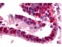
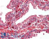
Polyclonal Rabbit anti‑Human UBA1 / UBE1 Antibody (IHC, WB) LS‑C18912
Polyclonal Rabbit anti‑Human UBA1 / UBE1 Antibody (IHC, WB) LS‑C18912
Antibody:
UBA1 / UBE1 Rabbit anti-Human Polyclonal Antibody
Application:
IHC, IHC-P, WB, ELISA
Reactivity:
Human
Format:
Unconjugated, Unmodified
Toll Free North America
 206-374-1102
For Research Use Only
Overview
Antibody:
UBA1 / UBE1 Rabbit anti-Human Polyclonal Antibody
Application:
IHC, IHC-P, WB, ELISA
Reactivity:
Human
Format:
Unconjugated, Unmodified
Specifications
Description
UBE1 antibody LS-C18912 is an unconjugated rabbit polyclonal antibody to human UBE1 (UBA1). Validated for ELISA, IHC and WB.
Target
Human UBA1 / UBE1
Synonyms
UBA1 | A1S9 | A1ST | A1S9T | AMCX1 | GXP1 | Protein A1S9 | SMAX2 | Ubiquitin-activating enzyme E1 | UBE1X | POC20 | UBE1 | UBA1A
Host
Rabbit
Reactivity
Human
(tested or 100% immunogen sequence identity)
Clonality
IgG
Polyclonal
Conjugations
Unconjugated
Purification
Protein A purified
Modifications
Unmodified
Immunogen
Anti-Ubiquitin Activating Enzyme E1 antibody was prepared from whole rabbit serum produced by repeated immunizations with a recombinant protein corresponding to full length Human Ubiquitin Activating Enzyme E1.
Applications
- IHC
- IHC - Paraffin (2 - 20 µg/ml)
- Western blot (1:1000 - 1:5000)
- ELISA (1:2000 - 1:10000)

|
Performing IHC? See our complete line of Immunohistochemistry Reagents including antigen retrieval solutions, blocking agents
ABC Detection Kits and polymers, biotinylated secondary antibodies, substrates and more.
|
Usage
This purified antibody has been tested for use in ELISA, immunohistochemistry and western blot. Specific conditions for reactivity should be optimized by the end user. Expect a band at ~118 kD in size corresponding to UBE1 by western blotting in the appropriate cell lysate or extract.
Presentation
Lyophilized from 0.02 M Potassium Phosphate, pH 7.2, 0.15 M NaCl, 0.01% Sodium Azide
Reconstitution
Reconstitute in 500 µL of deionized water.
Storage
Store vial at 4°C prior to restoration. Reconstitute with 0.1 ml of deionized water or equivalent. For extended storage aliquot contents and freeze at -20°C or below. Avoid freeze-thaw cycles.
Restrictions
For research use only. Intended for use by laboratory professionals.
About UBA1 / UBE1
Publications (0)
Customer Reviews (0)
Featured Products
Reactivity:
Mouse
Range:
31.25-2000 pg/ml
Species:
Human, Monkey, Mouse, Rat, Dog
Applications:
IHC, IHC - Paraffin, Immunofluorescence, Western blot

Request SDS/MSDS
To request an SDS/MSDS form for this product, please contact our Technical Support department at:
Technical.Support@LSBio.com
Requested From: United States
Date Requested: 4/17/2024
Date Requested: 4/17/2024


