Login
Registration enables users to use special features of this website, such as past
order histories, retained contact details for faster checkout, review submissions, and special promotions.
order histories, retained contact details for faster checkout, review submissions, and special promotions.
Forgot password?
Registration enables users to use special features of this website, such as past
order histories, retained contact details for faster checkout, review submissions, and special promotions.
order histories, retained contact details for faster checkout, review submissions, and special promotions.
Quick Order
Products
Antibodies
ELISA and Assay Kits
Research Areas
Infectious Disease
Resources
Purchasing
Reference Material
Contact Us
Locations
Orders Processing,
Shipping & Receiving,
Warehouse
2 Shaker Rd Suites
B001/B101
Shirley, MA 01464
Production Lab
Floor 6, Suite 620
20700 44th Avenue W
Lynnwood, WA 98036
Telephone Numbers
Tel: +1 (206) 374-1102
Fax: +1 (206) 577-4565
Contact Us
Additional Contact Details
Login
Registration enables users to use special features of this website, such as past
order histories, retained contact details for faster checkout, review submissions, and special promotions.
order histories, retained contact details for faster checkout, review submissions, and special promotions.
Forgot password?
Registration enables users to use special features of this website, such as past
order histories, retained contact details for faster checkout, review submissions, and special promotions.
order histories, retained contact details for faster checkout, review submissions, and special promotions.
Quick Order
| Catalog Number | Size | Price |
|---|---|---|
| LS-C96491-400 | 400 µl | $393 |
1 of 4
2 of 4
3 of 4
4 of 4
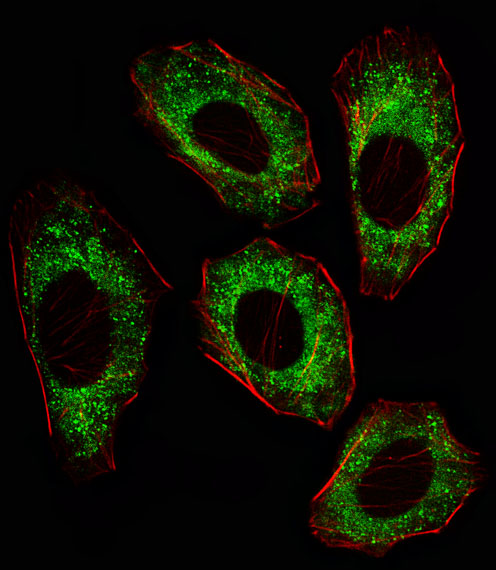
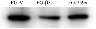
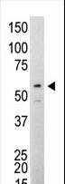
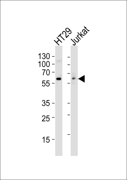
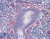
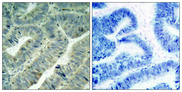
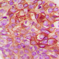
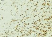
Monoclonal Mouse anti‑Human SRC Antibody (clone 17AT28, IF, WB) LS‑C96491
Monoclonal Mouse anti‑Human SRC Antibody (clone 17AT28, IF, WB) LS‑C96491

Antibody:
SRC Mouse anti-Human Monoclonal (17AT28) Antibody
Application:
IF, WB
Reactivity:
Human
Format:
Unconjugated, Unmodified
Toll Free North America
 206-374-1102
For Research Use Only
Overview
Antibody:
SRC Mouse anti-Human Monoclonal (17AT28) Antibody
Application:
IF, WB
Reactivity:
Human
Format:
Unconjugated, Unmodified
Specifications
Description
SRC antibody LS-C96491 is an unconjugated mouse monoclonal antibody to human SRC. Validated for IF and WB. Cited in 1 publication.
Target
Human SRC
Synonyms
SRC | ASV | Pp60c-src | Tyrosine kinase pp60c-src | Tyrosine-protein kinase SRC-1 | v-src | C-SRC | p60-Src | Proto-oncogene c-Src | SRC1
Host
Mouse
Reactivity
Human
(tested or 100% immunogen sequence identity)
Clonality
IgG1
Monoclonal
Clone
17AT28
Conjugations
Unconjugated
Purification
Protein G purified
Modifications
Unmodified
Applications
- Immunofluorescence (1:10 - 1:50)
- Western blot (1:100 - 1:500)
Presentation
PBS, 0.09% Sodium Azide
Storage
Maintain refrigerated at 2°C to 8°C for up to 6 months. For long term storage store at -20°C.
Restrictions
For research use only. Intended for use by laboratory professionals.
About SRC
LSBio Ratings
SRC Antibody (clone 17AT28) for IF/Immunofluorescence, WB/Western LS-C96491 has an LSBio Rating of
Publications (4)
Learn more about The LSBio Ratings Algorithm
Publications (1)
Interferon ß (IFN-ß) Production during the Double-stranded RNA (dsRNA) Response in Hepatocytes Involves Coordinated and Feedforward Signaling through Toll-like Receptor 3 (TLR3), RNA-dependent Protein Kinase (PKR), Inducible Nitric Oxide Synthase (iNOS), and Src Protein. Zhang L, Xiang W, Wang G, Yan Z, Zhu Z, Guo Z, Sengupta R, Chen AF, Loughran PA, Lu B, Wang Q, Billiar TR. The Journal of biological chemistry. 2016 291:15093-107. (WB; Human)
Customer Reviews (0)
Featured Products
Species:
Human, Mouse
Applications:
IHC, IHC - Paraffin, Immunofluorescence, Western blot
Species:
Human, Mouse, Rat, Dog
Applications:
Western blot, ELISA
Species:
Human, Mouse, Rat
Applications:
IHC, Western blot, Peptide Enzyme-Linked Immunosorbent Assay
Species:
Human, Mouse, Rat, Bovine, Pig, Chicken, Zebrafish
Applications:
IHC, IHC - Paraffin, ICC, Immunofluorescence, Western blot
Species:
Human, Monkey, Mouse, Rat
Applications:
IHC, Immunofluorescence, Western blot, Peptide Enzyme-Linked Immunosorbent Assay
Source:
E. coli
Tag:
6His, N-terminus
Request SDS/MSDS
To request an SDS/MSDS form for this product, please contact our Technical Support department at:
Technical.Support@LSBio.com
Requested From: United States
Date Requested: 4/24/2024
Date Requested: 4/24/2024


