Login
Registration enables users to use special features of this website, such as past
order histories, retained contact details for faster checkout, review submissions, and special promotions.
order histories, retained contact details for faster checkout, review submissions, and special promotions.
Forgot password?
Registration enables users to use special features of this website, such as past
order histories, retained contact details for faster checkout, review submissions, and special promotions.
order histories, retained contact details for faster checkout, review submissions, and special promotions.
Quick Order
Products
Antibodies
ELISA and Assay Kits
Research Areas
Infectious Disease
Resources
Purchasing
Reference Material
Contact Us
Locations
Orders Processing,
Shipping & Receiving,
Warehouse
2 Shaker Rd Suites
B001/B101
Shirley, MA 01464
Production Lab
Floor 6, Suite 620
20700 44th Avenue W
Lynnwood, WA 98036
Telephone Numbers
Tel: +1 (206) 374-1102
Fax: +1 (206) 577-4565
Contact Us
Additional Contact Details
Login
Registration enables users to use special features of this website, such as past
order histories, retained contact details for faster checkout, review submissions, and special promotions.
order histories, retained contact details for faster checkout, review submissions, and special promotions.
Forgot password?
Registration enables users to use special features of this website, such as past
order histories, retained contact details for faster checkout, review submissions, and special promotions.
order histories, retained contact details for faster checkout, review submissions, and special promotions.
Quick Order
| Catalog Number | Size | Price |
|---|---|---|
| LS-C745005-100 | 100 µg (1 mg/ml) | $567 |
1 of 8
2 of 8
3 of 8
4 of 8
5 of 8
6 of 8
7 of 8
8 of 8
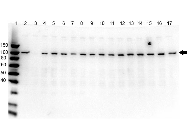
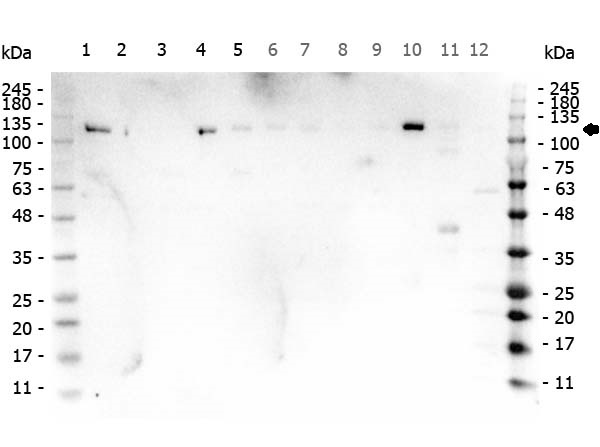
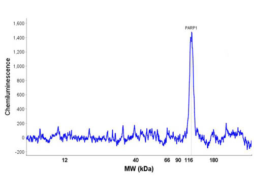
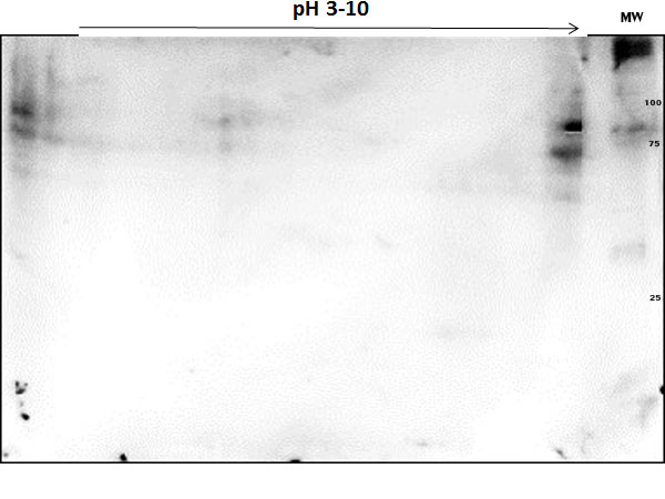
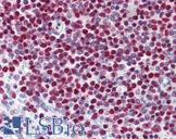
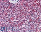
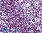
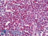
Polyclonal Rabbit anti‑Human PARP1 Antibody (N‑Terminus, IHC, WB) LS‑C745005
Polyclonal Rabbit anti‑Human PARP1 Antibody (N‑Terminus, IHC, WB) LS‑C745005

Antibody:
PARP1 Rabbit anti-Human Polyclonal (N-Terminus) Antibody
Application:
IHC, WB, IE
Reactivity:
Human
Format:
Unconjugated, Unmodified
Toll Free North America
 206-374-1102
For Research Use Only
Overview
Antibody:
PARP1 Rabbit anti-Human Polyclonal (N-Terminus) Antibody
Application:
IHC, WB, IE
Reactivity:
Human
Format:
Unconjugated, Unmodified
Specifications
Description
PARP1 antibody LS-C745005 is an unconjugated rabbit polyclonal antibody to human PARP1 (N-Terminus). Validated for IE, IHC and WB. Cited in 1 publication.
Target
Human PARP1
Synonyms
PARP1 | ADPRT | ADP-ribosyltransferase NAD(+) | Adp-ribosyltransferase | ADPRT1 | ARTD1 | ADPRT 1 | Poly(ADP-ribose) synthetase | Poly(ADP-ribosyl)transferase | PARP | Poly [ADP-ribose] polymerase 1 | PADPRT-1 | PARP-1 | Poly (ADP-ribose) polymerase 1 | Poly(ADP-ribose) polymerase | Poly[ADP-ribose] synthase 1 | PPOL
Host
Rabbit
Reactivity
Human
(tested or 100% immunogen sequence identity)
Clonality
IgG
Polyclonal
Conjugations
Unconjugated
Purification
Affinity chromatography
Modifications
Unmodified
Immunogen
PARP1 (N-term ZF1) purified antibody was prepared from whole rabbit serum produced by repeated immunizations with n-terminus region of human PARP1 zinc finger domain recombinant protein.
Epitope
N-Terminus
Specificity
This antibody is specific for human PARP1 protein. No cross reactivity detected towards other PARP members when using siRNAs against 18 PARP family members. Cross-reactivity with PARP1 from other sources has not been determined.
Applications
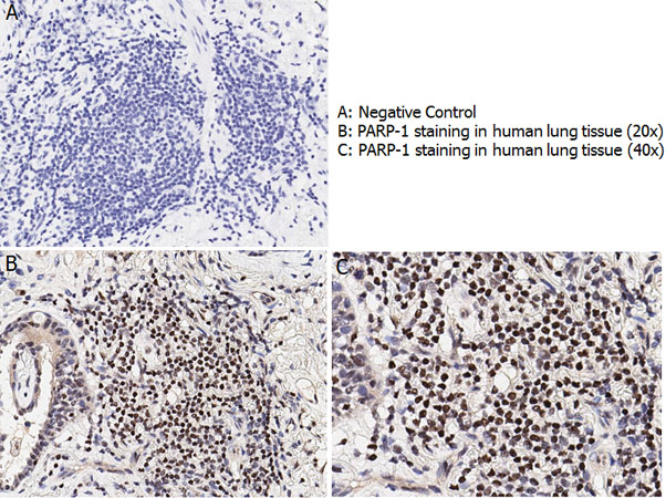
- IHC (1:100)
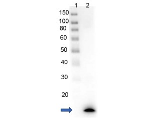
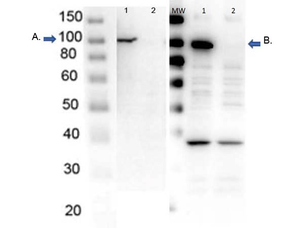
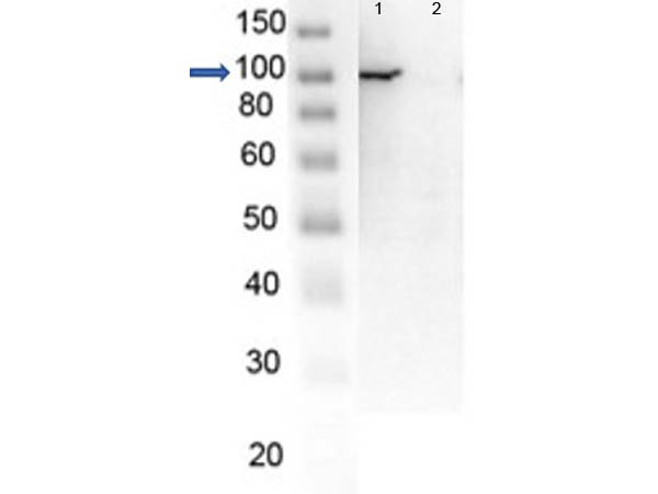
- Western blot (1:1000)
- Immunoelectrophoresis

|
Performing IHC? See our complete line of Immunohistochemistry Reagents including antigen retrieval solutions, blocking agents
ABC Detection Kits and polymers, biotinylated secondary antibodies, substrates and more.
|
Usage
Applications should be user optimized.
Presentation
0.02 M Potassium Phosphate, pH 7.2, 0.15 M NaCl, 0.01% Sodium Azide
Storage
Store vial at -20°C or below prior to opening. Dilute 1:10 to minimize loss. Store the vial at -20°C or below after dilution. Avoid freeze-thaw cycles.
Restrictions
For research use only. Intended for use by laboratory professionals.
About PARP1
LSBio Ratings
PARP1 Antibody (N-Terminus) for IHC, WB/Western LS-C745005 has an LSBio Rating of
Publications (4)
Learn more about The LSBio Ratings Algorithm
Publications (1)
Tracking the expression of therapeutic protein targets in rare cells by antibody-mediated nanoparticle labelling and magnetic sorting. Mahmoud Labib, Zongjie Wang, Sharif U Ahmed, Reza M Mohamadi, Bill Duong, Brenda Green, Edward H Sargent, Shana O Kelley. Nature biomedical engineering. 2021 January;5:41-52.
Customer Reviews (0)
Featured Products
Species:
Human
Applications:
IHC, IHC - Paraffin, IHC - Frozen, Immunofluorescence, Western blot, Immunoprecipitation, ELISA
Species:
Human, Mouse, Rat
Applications:
IHC, IHC - Paraffin, Western blot
Species:
Human, Monkey, Mouse, Rat
Applications:
IHC, IHC - Paraffin, Immunofluorescence, Western blot, Immunoprecipitation, ELISA
Species:
Human, Mouse, Bovine, Chicken, Xenopus
Applications:
IHC, IHC - Paraffin, IHC - Frozen, ICC, Western blot, Immunoprecipitation, ELISA

Request SDS/MSDS
To request an SDS/MSDS form for this product, please contact our Technical Support department at:
Technical.Support@LSBio.com
Requested From: United States
Date Requested: 4/16/2024
Date Requested: 4/16/2024


