Login
Registration enables users to use special features of this website, such as past
order histories, retained contact details for faster checkout, review submissions, and special promotions.
order histories, retained contact details for faster checkout, review submissions, and special promotions.
Forgot password?
Registration enables users to use special features of this website, such as past
order histories, retained contact details for faster checkout, review submissions, and special promotions.
order histories, retained contact details for faster checkout, review submissions, and special promotions.
Quick Order
Products
Antibodies
ELISA and Assay Kits
Research Areas
Infectious Disease
Resources
Purchasing
Reference Material
Contact Us
Locations
Orders Processing,
Shipping & Receiving,
Warehouse
2 Shaker Rd Suites
B001/B101
Shirley, MA 01464
Production Lab
Floor 6, Suite 620
20700 44th Avenue W
Lynnwood, WA 98036
Telephone Numbers
Tel: +1 (206) 374-1102
Fax: +1 (206) 577-4565
Contact Us
Additional Contact Details
Login
Registration enables users to use special features of this website, such as past
order histories, retained contact details for faster checkout, review submissions, and special promotions.
order histories, retained contact details for faster checkout, review submissions, and special promotions.
Forgot password?
Registration enables users to use special features of this website, such as past
order histories, retained contact details for faster checkout, review submissions, and special promotions.
order histories, retained contact details for faster checkout, review submissions, and special promotions.
Quick Order
| Catalog Number | Size | Price |
|---|---|---|
| LS-C313066-10 | 10 µg | $318 |
| LS-C313066-100 | 100 µg | $470 |
1 of 4
2 of 4
3 of 4
4 of 4
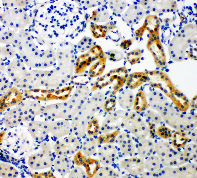
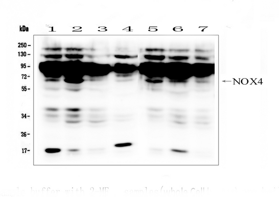
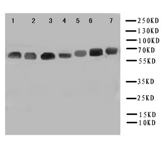
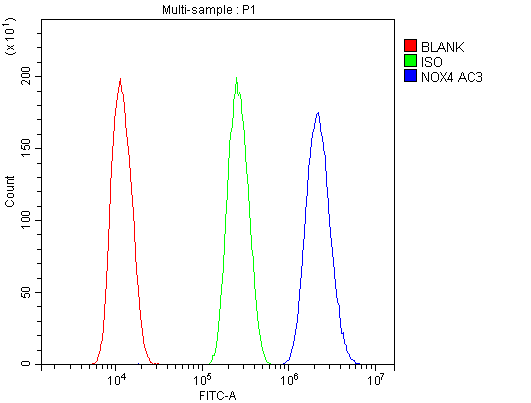
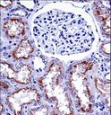
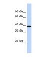
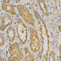
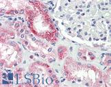
Polyclonal Rabbit anti‑Mouse NOX4 Antibody (aa561‑578, IHC, WB) LS‑C313066
Polyclonal Rabbit anti‑Mouse NOX4 Antibody (aa561‑578, IHC, WB) LS‑C313066

Note: This antibody replaces LS-C388976
Antibody:
NOX4 Rabbit anti-Mouse Polyclonal (aa561-578) Antibody
Application:
IHC, IHC-P, WB
Reactivity:
Mouse, Human, Rat
Format:
Unconjugated, Unmodified
Toll Free North America
 206-374-1102
For Research Use Only
Overview
Antibody:
NOX4 Rabbit anti-Mouse Polyclonal (aa561-578) Antibody
Application:
IHC, IHC-P, WB
Reactivity:
Mouse, Human, Rat
Format:
Unconjugated, Unmodified
Specifications
Description
NOX4 antibody LS-C313066 is an unconjugated rabbit polyclonal antibody to NOX4 (aa561-578) from mouse. It is reactive with human, mouse and rat. Validated for IHC and WB. Cited in 2 publications.
Target
Mouse NOX4
Synonyms
NOX4 | Kidney oxidase-1 | KOX | RENOX | KOX-1 | NADPH oxidase 4 | Renal NAD(P)H-oxidase
Host
Rabbit
Reactivity
Mouse, Human, Rat
(tested or 100% immunogen sequence identity)
Clonality
Polyclonal
Conjugations
Unconjugated
Purification
Immunogen affinity purified
Modifications
Unmodified
Immunogen
A synthetic peptide corresponding to a sequence at the C-Terminus of mouse NADPH oxidase 4(561-578 aa NRNNSYGTKFEYNKES), identical to the related rat sequence and different from the related human sequence by two amino acids.
Epitope
aa561-578
Specificity
EXpressed in brain, in all layers of the cerebellum, in pyramidal cells of the Ammon horn and in Purkinje cells (at protein level). Expressed in osteoclasts, leukocytes, kidney, liver and lung. .
Applications
- IHC
- IHC - Paraffin (0.5 - 1 µg/ml)
- Western blot (0.1 - 0.5 µg/ml)

|
Performing IHC? See our complete line of Immunohistochemistry Reagents including antigen retrieval solutions, blocking agents
ABC Detection Kits and polymers, biotinylated secondary antibodies, substrates and more.
|
Presentation
Lyophilized from 0.2mg Na2HPO4, 5mg BSA, 0.9mg NaCl, 0.05mg Thimerosal, 0.05mg sodium azide.
Reconstitution
Add 0.2ml of distilled water will yield a concentration of 500µg/ml.
Storage
At -20°C for 1 year. After reconstitution, at 4°C for 1 month. It can also be aliquotted and stored frozen at -20°C for a longer time. Avoid freeze-thaw cycles.
Restrictions
For research use only. Intended for use by laboratory professionals.
About NOX4
LSBio Ratings
NOX4 Antibody (aa561-578) for IHC, WB/Western LS-C313066 has an LSBio Rating of
Publications (4.1)
Learn more about The LSBio Ratings Algorithm
Publications (2)
Nuclear NADPH oxidase-4 associated with disease progression in renal cell carcinoma. Dharam Kaushik, Keith A Ashcraft, Hanzhang Wang, Karthigayan Shanmugasundaram, Pankil K Shah, Gabriela Gonzalez, Alia Nazarullah, Cooper B Tye, Michael A Liss, Deepak K Pruthi, Ahmed M Mansour, Wasim Chowdhury, Dean Bacich, Hao Zhang, Amanda L Watson, Karen Block, Denise O'Keefe, Ronald Rodriguez. Translational research : the journal of laboratory and clinical medicine. 2020 Sep;;223:44575.
Effects of anagliptin on the stress induced accelerated senescence of human umbilical vein endothelial cells. Seon Mee Kang , Hye Sook Jung, Min Jeong Kwon, Soon Hee Lee, Jeong Hyun Park. Annals of Translational Medicine. 2021 May;;9:750.
Customer Reviews (0)
Featured Products
Species:
Human, Mouse, Rat, Bovine, Sheep, Primate
Applications:
IHC, IHC - Paraffin, Western blot
Request SDS/MSDS
To request an SDS/MSDS form for this product, please contact our Technical Support department at:
Technical.Support@LSBio.com
Requested From: United States
Date Requested: 4/18/2024
Date Requested: 4/18/2024


