Products
Research Areas
COVID-19
Resources
Login
Quick Order
Cart
Login
Registration enables users to use special features of this website, such as past
order histories, retained contact details for faster checkout, review submissions, and special promotions.


Fields marked with a * are required.

Login
Quick Order
Contact Us

Locations


Orders Processing,
Shipping & Receiving,
Warehouse

2 Shaker Rd Suites
B001/B101
Shirley, MA 01464


Production Lab

Floor 6, Suite 620
20700 44th Avenue W
Lynnwood, WA 98036

Telephone Numbers


Tel: +1 (206) 374-1102
Fax: +1 (206) 577-4565

Contact Us


Additional Contact Details

Login
Registration enables users to use special features of this website, such as past
order histories, retained contact details for faster checkout, review submissions, and special promotions.


Fields marked with a * are required.

Login
Quick Order
Catalog Number Size Price
LS-C744815-25 25 µl (1 mg/ml) $304 
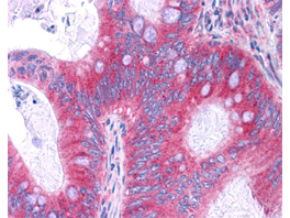
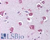
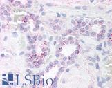
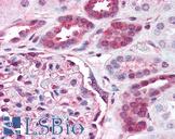
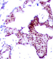
RELA / NFKB p65 Antibody - Antibody 200-301-065 has been tested in immunohistochemistry, analyzed by an anatomic pathologist and validated for use in IHC applications against formalin-fixed, paraffin-embedded human tissues. showed moderate to strong staining within many tissues, including epithelium of the breast, small intestine, kidney, pancreas, prostate, skin, placenta, and uterus, as well as within neurons and lymphoid tissues such as spleen, thymus, and tonsil. The antibody produced an excellent signal with almost no background staining at a concentration of 2.5 ug/ml. The image displayed shows specific staining in colon carcinoma as the precipitated red signal, with a hematoxylin purple nuclear counterstain.
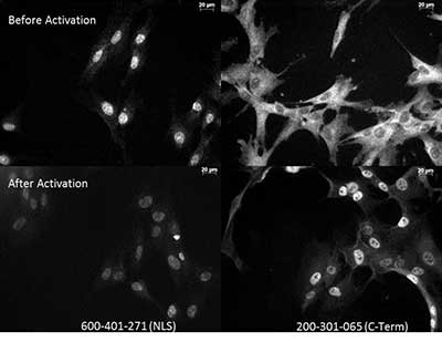
RELA / NFKB p65 Antibody - Anti NFkB monoclonal antibody – Immunocytochemistry Tissue: Human Fibroblasts Top: Before activation Bottom: After activation with poly IC Left: 600-401-271 anti p65 NLS specific lot 18372 Right: 200-301-065 Monoclonal antibody C-Term The two antibodies that are shown target different regions of the p65 protein. The different staining patterns are thought to correspond with different functional regions of the protein.
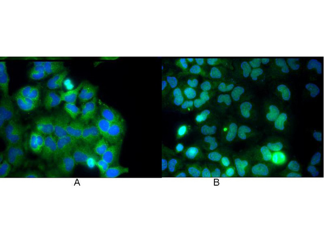
RELA / NFKB p65 Antibody - Monoclonal anti NFKB p65 (Rel A) antibody was used to detect p65 by immunofluorescence at a dilution of 1:5000. Hela cells were grown to sub-confluent on 18 mm2 glass coverslips #1.5. Cells were either unstimulated (A), or stimulated (B) with 50 ng/ml of TNF alpha for 30 min prior fixation. Cells were then fixed in methanol and blocked with 10% normal goat serum (NGS), in PBS, and TritonX 0.2% (Tx) and incubated for 1 hr at RT with primary ab, counterstained with DAPI and washed in PBS/NGS/Tx. Cells were incubated for 1 hr at RT with Atto 425 conjugated anti mouse secondary antibody for STED CW imaging. Data was collected on a STED-CW TCS-SP5 Confocal system equipped with a DFC 350FX camera allowing sequential acquisition in widefiled, confocal and STED CW imaging on the same system.
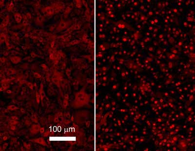
RELA / NFKB p65 Antibody - Monoclonal anti NFKB p65 (Rel A) antibody was used to detect p65 in b.end5 mouse endothelial cells. Unstimulated control cells (left) show cytoplasmic staining, TNF-alpha stimulated cells (right) show nuclear staining. For staining, cells were washed with PBS to remove all traces of culture media and fixed with paraformaldehyde 4 % (45 min). Slides were washed with PBT (phosphate buffer 0.1M + Triton-X-100 0.1 %), 3 times 10 min each, then, blocked with PBT + 5 % normal goat serum, 1 hour. Sample was Incubated overnight in primary antibody. After 3X wash in PBT for 10 min, slides were incubated 1 hour with secondary antibody and mounted in 1:1 PB glycerol.
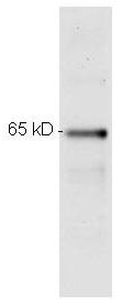
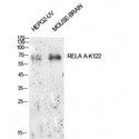
RELA / NFKB p65 Antibody - Monoclonal anti p65 200-301-065 was used to detect p65 by Western blot. Samples were prepared in RIPA lysis buffer, boiled with NuPage 4x LDS Sample Buffer and run on NuPage 4-12% Bis-Tris Gels. Blot was incubated with primary antibody at a dilution of 1:500 and detected with HRP conjugated anti mouse antibody at a dilution of 1:10000.
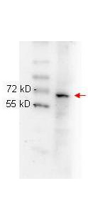
RELA / NFKB p65 Antibody - Anti NFKB p65 (Rel A) monoclonal antibody 200-301-065 was used to detect ~65 kD band (red arrow) in HeLa whole cell lysate. Lysate was run on 4-20% gradient gel transferred under standard conditions and blocked in 1% BSA-TTBS 30 min RT. Blot was probed with monoclonal anti p65 1:1000 in 1% BSA-TBS-T o/n 4°C and detected with HRP conjugated Rb-anti Mouse antibody 1:40,000 in MB-070 30 min RT.
1 of 6
2 of 6
3 of 6
4 of 6
5 of 6
6 of 6

Monoclonal Mouse anti‑Human RELA / NFKB p65 Antibody (clone 27F9.G4, C‑Terminus, IHC, IF, WB) LS‑C744815

Monoclonal Mouse anti‑Human RELA / NFKB p65 Antibody (clone 27F9.G4, C‑Terminus, IHC, IF, WB) LS‑C744815

Antibody:
RELA / NFKB p65 Mouse anti-Human Monoclonal (C-Terminus) (27F9.G4) Antibody
Application:
IHC, ICC, IF, WB, ELISA
Reactivity:
Human
Format:
Unconjugated, Unmodified
Price
Catalog Number
$304
LS-C744815-25
Toll Free North America
206-374-1102
For Research Use Only

Overview

Antibody:
RELA / NFKB p65 Mouse anti-Human Monoclonal (C-Terminus) (27F9.G4) Antibody
Application:
IHC, ICC, IF, WB, ELISA
Reactivity:
Human
Format:
Unconjugated, Unmodified

Specifications

Description
NFKB p65 antibody LS-C744815 is an unconjugated mouse monoclonal antibody to human NFKB p65 (RELA) (C-Terminus). Validated for ELISA, ICC, IF, IHC and WB.
Target
Human RELA / NFKB p65
Synonyms
RELA | NF-kappa-B p65delta3 | NF-kappaB | NFKB3 | p65 | Transcription factor p65 | NF kappaB | NFKB
Host
Mouse
Reactivity
Human (tested or 100% immunogen sequence identity)
Clonality
IgG2a,k Monoclonal
Clone
27F9.G4
Conjugations
Unconjugated
Purification
Protein A affinity chromatography
Modifications
Unmodified
Immunogen
NFkB p65 (Rel A) peptide corresponding to a region near the C-terminus of the human protein conjugated to Keyhole Limpet Hemocyanin (KLH).
Epitope
C-Terminus
Applications
  • IHC (1:200 - 1:600)
  • ICC
  • Immunofluorescence (1:5000)
  • Western blot (1:1000 - 1:5000)
  • ELISA (1:50000 - 1:100000)
Performing IHC? See our complete line of Immunohistochemistry Reagents including antigen retrieval solutions, blocking agents ABC Detection Kits and polymers, biotinylated secondary antibodies, substrates and more.
Usage
Applications should be user optimized.
Presentation
0.02 M Potassium Phosphate, pH 7.2, 0.15 M NaCl, 0.01% Sodium Azide
Storage
Store vial at -20°C or below prior to opening. Dilute 1:10 to minimize loss. Store the vial at -20°C or below after dilution. Avoid freeze-thaw cycles.
Restrictions
For research use only. Intended for use by laboratory professionals.
Guarantee
This antibody carries the LSBio 100% Guarantee.
LSBio Guarantee
About RELA / NFKB p65
Q04206 NM_021975 NP_068810.3

Publications (0)

Customer Reviews (0)


Request SDS/MSDS

To request an SDS/MSDS form for this product, please contact our Technical Support department at:

Technical.Support@LSBio.com

Requested From: United States
Date Requested: 4/19/2024