Login
Registration enables users to use special features of this website, such as past
order histories, retained contact details for faster checkout, review submissions, and special promotions.
order histories, retained contact details for faster checkout, review submissions, and special promotions.
Forgot password?
Registration enables users to use special features of this website, such as past
order histories, retained contact details for faster checkout, review submissions, and special promotions.
order histories, retained contact details for faster checkout, review submissions, and special promotions.
Quick Order
Products
Antibodies
ELISA and Assay Kits
Research Areas
Infectious Disease
Resources
Purchasing
Reference Material
Contact Us
Locations
Orders Processing,
Shipping & Receiving,
Warehouse
2 Shaker Rd Suites
B001/B101
Shirley, MA 01464
Production Lab
Floor 6, Suite 620
20700 44th Avenue W
Lynnwood, WA 98036
Telephone Numbers
Tel: +1 (206) 374-1102
Fax: +1 (206) 577-4565
Contact Us
Additional Contact Details
Login
Registration enables users to use special features of this website, such as past
order histories, retained contact details for faster checkout, review submissions, and special promotions.
order histories, retained contact details for faster checkout, review submissions, and special promotions.
Forgot password?
Registration enables users to use special features of this website, such as past
order histories, retained contact details for faster checkout, review submissions, and special promotions.
order histories, retained contact details for faster checkout, review submissions, and special promotions.
Quick Order
| Catalog Number | Size | Price |
|---|---|---|
| LS-B2365-50 | 50 µg (0.5 mg/ml) | $485 |

1 of 6
2 of 6
3 of 6
4 of 6
5 of 6
6 of 6
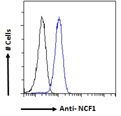
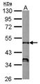
IHC‑plus™ Polyclonal Goat anti‑Human NCF1 / p47phox / p47 phox Antibody (aa378‑390, IHC, IF, WB) LS‑B2365
IHC‑plus™ Polyclonal Goat anti‑Human NCF1 / p47phox / p47 phox Antibody (aa378‑390, IHC, IF, WB) LS‑B2365

Note: This antibody replaces LS-C20183, LS-C9424, LS-C91063
Antibody:
NCF1 / p47phox / p47 phox Goat anti-Human Polyclonal (aa378-390) Antibody
Application:
IHC, IHC-P, IF, WB, Peptide-ELISA
Reactivity:
Human, Monkey, Bovine
Format:
Unconjugated, Unmodified
Toll Free North America
 206-374-1102
For Research Use Only
Overview
Antibody:
NCF1 / p47phox / p47 phox Goat anti-Human Polyclonal (aa378-390) Antibody
Application:
IHC, IHC-P, IF, WB, Peptide-ELISA
Reactivity:
Human, Monkey, Bovine
Format:
Unconjugated, Unmodified
Specifications
Description
P47 phox antibody LS-B2365 is an unconjugated goat polyclonal antibody to p47 phox (NCF1 / p47phox) (aa378-390) from human. It is reactive with human, bovine and monkey. Validated for IF, IHC, Peptide-ELISA and WB. Tested on 20 paraffin-embedded human tissues. Cited in 2 publications.
Target
Human NCF1 / p47phox / p47 phox
Synonyms
NCF1 | NCF1A | Neutrophil cytosolic factor 1 | NCF-47K | Nox organizer 2 | NOXO2 | p47-phox | p47phox | SH3PXD1A | NADPH oxidase organizer 2 | NCF-1 | Neutrophil cytosol factor 1 | Nox-organizing protein 2
Host
Goat
Reactivity
Human, Monkey, Bovine
(tested or 100% immunogen sequence identity)
Clonality
Polyclonal
Conjugations
Unconjugated
Purification
Purified from goat serum by ammonium sulphate precipitation followed by antigen affinity chromatography using the immunizing peptide.
Modifications
Unmodified
Immunogen
Peptide with sequence C-SESTKRKLASAV, from the C-Terminus of protein sequence according to NP_000256.4.
Epitope
aa378-390
Specificity
Human NCF1 / p47phox.
Applications
- IHC
- IHC - Paraffin (5 µg/ml)
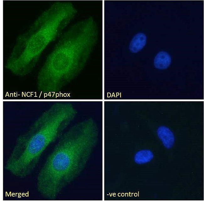
- Immunofluorescence (10 µg/ml)
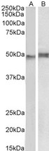
- Western blot (0.01 - 1 µg/ml)
- Peptide Enzyme-Linked Immunosorbent Assay (1:20000)

|
Performing IHC? See our complete line of Immunohistochemistry Reagents including antigen retrieval solutions, blocking agents
ABC Detection Kits and polymers, biotinylated secondary antibodies, substrates and more.
|
Usage
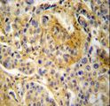
Immunohistochemistry: LS-B2365 was validated for use in immunohistochemistry on a panel of 21 formalin-fixed, paraffin-embedded (FFPE) human tissues after heat induced antigen retrieval in pH 6.0 citrate buffer. After incubation with the primary antibody, slides were incubated with biotinylated secondary antibody, followed by alkaline phosphatase-streptavidin and chromogen. The stained slides were evaluated by a pathologist to confirm staining specificity. The optimal working concentration for LS-B2365 was determined to be 5 ug/ml.
Presentation
TBS, pH 7.3, 0.02% Sodium Azide, 0.5% BSA
Storage
Aliquot and store at -20°C. Avoid freeze-thaw cycles.
Restrictions
For research use only. Intended for use by laboratory professionals.
About NCF1 / p47phox / p47 phox
Validation
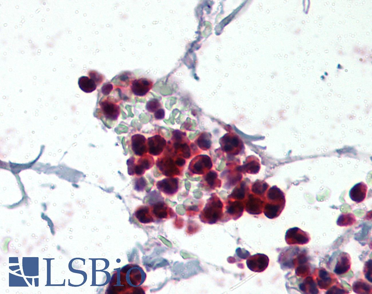
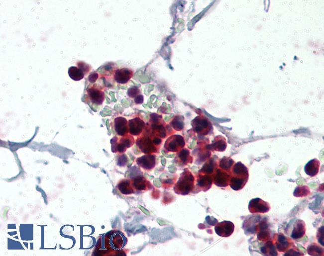
Anti-p47phox antibody IHC of human colon, neutrophils. Immunohistochemistry of formalin-fixed, paraffin-embedded tissue after heat-induced antigen retrieval. Antibody concentration 5 ug/ml.
Anti-p47phox antibody IHC of human colon, neutrophils. Immunohistochemistry of formalin-fixed, paraffin-embedded tissue after heat-induced antigen retrieval. Antibody concentration 5 ug/ml.
See More About...
LSBio Ratings
IHC-plus™ NCF1 / p47phox / p47 phox Antibody (aa378-390) for IHC, IF/Immunofluorescence, WB/Western LS-B2365 has an LSBio Rating of
Publications (4.1)
Laboratory Validation Score (4)
Learn more about The LSBio Ratings Algorithm
Publications (2)
NADPH oxidase expression in active multiple sclerosis lesions in relation to oxidative tissue damage and mitochondrial injury. Fischer MT, Sharma R, Lim JL, Haider L, Frischer JM, Drexhage J, Mahad D, Bradl M, van Horssen J, Lassmann H. Brain : a journal of neurology. 2012 135:886-99. (IHC-P; Human)
Characterization of the inflammatory response to solid cancer metastases in the human brain. Berghoff AS, Lassmann H, Preusser M, H[Character f6]ftberger R. Clinical & experimental metastasis. 2013 Jan;30:69-81.
Customer Reviews (0)
Featured Products
Species:
Human
Applications:
IHC, Western blot, ELISA
Species:
Human, Bovine
Applications:
ICC, Western blot, Immunoprecipitation

Species:
Human, Mouse, Rat
Applications:
IHC, IHC - Paraffin, Immunofluorescence, Peptide Enzyme-Linked Immunosorbent Assay
Species:
Human
Applications:
IHC, IHC - Paraffin, Western blot
Reactivity:
Human
Range:
0.312-20 ng/ml
Request SDS/MSDS
To request an SDS/MSDS form for this product, please contact our Technical Support department at:
Technical.Support@LSBio.com
Requested From: United States
Date Requested: 4/19/2024
Date Requested: 4/19/2024


