Products
Research Areas
COVID-19
Resources
Login
Quick Order
Cart
Login
Registration enables users to use special features of this website, such as past
order histories, retained contact details for faster checkout, review submissions, and special promotions.


Fields marked with a * are required.

Login
Quick Order
Contact Us

Locations


Orders Processing,
Shipping & Receiving,
Warehouse

2 Shaker Rd Suites
B001/B101
Shirley, MA 01464


Production Lab

Floor 6, Suite 620
20700 44th Avenue W
Lynnwood, WA 98036

Telephone Numbers


Tel: +1 (206) 374-1102
Fax: +1 (206) 577-4565

Contact Us


Additional Contact Details

Login
Registration enables users to use special features of this website, such as past
order histories, retained contact details for faster checkout, review submissions, and special promotions.


Fields marked with a * are required.

Login
Quick Order
Catalog Number Size Price
LS-C745286-25 25 µl (1 mg/ml) $304 
MYL12A / MRCL3 Antibody - Affinity purified anti-Monophosphorylated RLC Smooth and Non-Muscle Myosin pS19/20 antibody was used at 2.5 µg/ml to detect signal in a variety of tissues including multi-human, multi-brain and multi-cancer slides. This image shows strong staining of both vascular and myometrial smooth muscle cells of the uterus. Tissue was formalin-fixed and paraffin embedded. The image shows localization of the antibody as the precipitated red signal, with a hematoxylin purple nuclear counterstain.
MYL12A / MRCL3 Antibody - Immunohistochemistry with anti-myosin pS19/pS20 antibody showing strong cytoplasmic staining of myocytes in mouse heart muscle 20x and 40x (B & C). Staining was performed on Leica Bond system using the standard protocol. Formalin fixed/paraffin embedded tissue sections were subjected to antigen retrieval and then incubated with rabbit anti-myosin pS19/pS20 antibody at 1:100 dilution for 60 minutes. Biotinylated Anti-rabbit secondary antibody was used to detect primary antibody. The reaction was developed using streptavidin-HRP conjugated compact polymer system and visualized with chromogen substrate, 3’3-diamino-benzidine substrate (DAB). The sections were then counterstained with hematoxylin to detect cell nuclei.
MYL12A / MRCL3 Antibody - Affinity Purified Phospho specific antibody to Monophosphorylated Regulatory Light Chain of Smooth and Non-muscle Myosin at pS19/pS20 was used at a 1:5000 dilution to detect myosin light chain by Western blot. Either 13 or 20 µl of a mouse cardiac myocyte lysate was loaded on a 4-20% Criterion gel for SDS-PAGE. Samples were either mock-treated or CLA-treated, as indicated. After washing, a 1:5,000 dilution of HRP conjugated Gt-a-Rabbit IgG preceded color development using Amersham's substrate system. Other detection methods will yield similar results. Data courtesy of the Alliance for Cellular Signaling (http://www.signaling-gateway.org).
MYL12A / MRCL3 Antibody - Western blot of rabbit Anti-Myosin pS19/pS20 primary antibody. Lane 1: Regulatory Light Chain Non-Phospho recombinant protein. Lane 2: Regulatory Light Chain Phospho recombinant protein. Lane 3: Smooth Muscle Non-Phospho recombinant protein. Lane 4: Smooth Muscle Phospho recombinant protein. Load: 50 ng per lane. Primary antibody: Myosin pS19/pS20 primary antibody at 1:1,000 overnight at 4°C. Secondary antibody: Peroxidase rabbit secondary antibody at 1:40,000 for 60 min at RT. Blocking: MB-070 for 30 min at RT. Predicted/Observed size: 20 kDa, 20 kDa for Regulatory Light Chain Phospho. Other band(s): None.
MYL12A / MRCL3 Antibody - Affinity purified phosphospecific antibody to phosphorylated regulatory light chain of smooth and non-muscle Myosin at pS19/pS20 was used at a 1:1000 dilution to detect myosin light chain by Western blot on 3T3 cell lysates. A standard urea/glycerol gel without SDS was used to separate phospho forms of regulatory light chain according to mass to charge ratios. In Panel A, reactivity of phosphospecific antibody is shown. In Panel B, reactivity of commercially available pan reactive antibody that detects both unphosphorylated and phosphorylated forms of regulatory light chain is shown. phosphospecific antibody detects both monophosphorylated (pSer20 Mono-P-RLC) and diphosphorylated (pThr19-pSer20 Di-P-RLC) regulatory light chain.
1 of 5
2 of 5
3 of 5
4 of 5
5 of 5

Polyclonal Rabbit anti‑Human MYL12A / MRCL3 Antibody (phospho‑Ser19/20, IHC, WB) LS‑C745286

Polyclonal Rabbit anti‑Human MYL12A / MRCL3 Antibody (phospho‑Ser19/20, IHC, WB) LS‑C745286

Antibody:
MYL12A / MRCL3 Rabbit anti-Human Polyclonal (pSer19/20) Antibody
Application:
IHC, WB, IP, ELISA
Reactivity:
Human
Format:
Unconjugated, Unmodified
Price
Catalog Number
$304
LS-C745286-25
Toll Free North America
206-374-1102
For Research Use Only

Overview

Antibody:
MYL12A / MRCL3 Rabbit anti-Human Polyclonal (pSer19/20) Antibody
Application:
IHC, WB, IP, ELISA
Reactivity:
Human
Format:
Unconjugated, Unmodified

Specifications

Description
MRCL3 antibody LS-C745286 is an unconjugated rabbit polyclonal antibody to human MRCL3 (MYL12A) (pSer19/20). Validated for ELISA, IHC, IP and WB.
Target
Human MYL12A / MRCL3
Synonyms
MYL12A | MRCL3 | MLCB | MRLC3 | MYL2B | MLC-2B
Host
Rabbit
Reactivity
Human (tested or 100% immunogen sequence identity)
Clonality
IgG Polyclonal
Conjugations
Unconjugated
Purification
Affinity purified
Modifications
Unmodified
Immunogen
Human Myosin Light Chain phospho peptide corresponding to a region near the amino terminus of the human smooth/non-muscle form of myosin regulatory light chain conjugated to Keyhole Limpet Hemocyanin (KLH).
Epitope
pSer19/20
Specificity
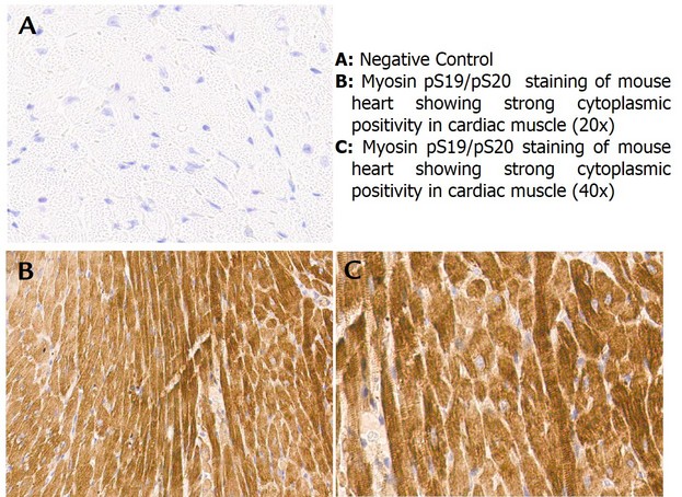
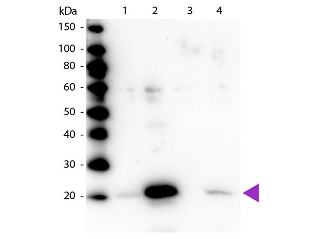
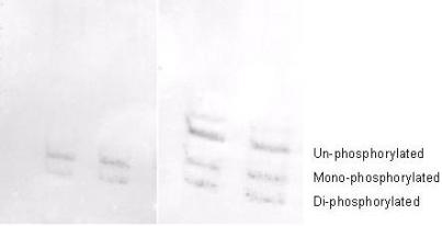
The antibody is phosphospecific and detects monophosphorylated and diphosphorylated forms of the protein. The product was affinity purified from monospecific antiserum by immunoaffinity purification. Antiserum was first purified against the phosphorylated form of the immunizing peptide. The resultant affinity purified antibody was then cross-adsorbed against the non-phosphorylated form of the immunizing peptide. This phosphospecific polyclonal antibody is specific for the phosphorylated pS19/pS20 form of the protein, depending on the source origin of the protein. Reactivity with non-phosphorylated myosin light chain is less than 1% by ELISA. Cross reactivity is expected with myosin light chain from human and mouse. Reactivity with the protein from other species has not been determined. However, the sequence of the immunogen is nearly identical in mammalian and avian species. BLAST search analysis was used to determine that the smooth and non-muscle forms of myosin regulatory light chain have identical sequences. Cross reactivity is expected.
Applications
  • IHC (2.5 µg/ml)
  • Western blot (1:1000 - 1:5000)
  • Immunoprecipitation (1:100)
  • ELISA (1:10000 - 1:30000)
Performing IHC? See our complete line of Immunohistochemistry Reagents including antigen retrieval solutions, blocking agents ABC Detection Kits and polymers, biotinylated secondary antibodies, substrates and more.
Usage
Applications should be user optimized.
Presentation
0.02 M Potassium Phosphate, pH 7.2, 0.15 M NaCl, 0.01% Sodium Azide
Storage
Store vial at -20°C or below prior to opening. Dilute 1:10 to minimize loss. Store the vial at -20°C or below after dilution. Avoid freeze-thaw cycles.
Restrictions
For research use only. Intended for use by laboratory professionals.
Guarantee
This antibody carries the LSBio 100% Guarantee.
LSBio Guarantee
About MYL12A / MRCL3
P19105 NM_006471 NP_006462.1

Publications (0)

Customer Reviews (0)


Request SDS/MSDS

To request an SDS/MSDS form for this product, please contact our Technical Support department at:

Technical.Support@LSBio.com

Requested From: United States
Date Requested: 4/19/2024