Login
Registration enables users to use special features of this website, such as past
order histories, retained contact details for faster checkout, review submissions, and special promotions.
order histories, retained contact details for faster checkout, review submissions, and special promotions.
Forgot password?
Registration enables users to use special features of this website, such as past
order histories, retained contact details for faster checkout, review submissions, and special promotions.
order histories, retained contact details for faster checkout, review submissions, and special promotions.
Quick Order
Products
Antibodies
ELISA and Assay Kits
Research Areas
Infectious Disease
Resources
Purchasing
Reference Material
Contact Us
Locations
Orders Processing,
Shipping & Receiving,
Warehouse
2 Shaker Rd Suites
B001/B101
Shirley, MA 01464
Production Lab
Floor 6, Suite 620
20700 44th Avenue W
Lynnwood, WA 98036
Telephone Numbers
Tel: +1 (206) 374-1102
Fax: +1 (206) 577-4565
Contact Us
Additional Contact Details
Login
Registration enables users to use special features of this website, such as past
order histories, retained contact details for faster checkout, review submissions, and special promotions.
order histories, retained contact details for faster checkout, review submissions, and special promotions.
Forgot password?
Registration enables users to use special features of this website, such as past
order histories, retained contact details for faster checkout, review submissions, and special promotions.
order histories, retained contact details for faster checkout, review submissions, and special promotions.
Quick Order
| Catalog Number | Size | Price |
|---|---|---|
| LS-C204825-100 | 100 µl (1 mg/ml) | $376 |
1 of 2
2 of 2
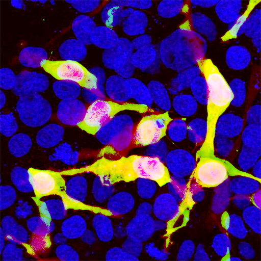
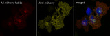
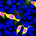
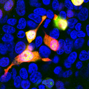
Polyclonal Chicken anti‑Discosoma mCherry Antibody (IF, WB) LS‑C204825
Polyclonal Chicken anti‑Discosoma mCherry Antibody (IF, WB) LS‑C204825

Antibody:
mCherry Chicken anti-Discosoma Polyclonal Antibody
Application:
ICC, IF, WB
Format:
Unconjugated, Unmodified
Toll Free North America
 206-374-1102
For Research Use Only
Overview
Antibody:
mCherry Chicken anti-Discosoma Polyclonal Antibody
Application:
ICC, IF, WB
Format:
Unconjugated, Unmodified
Specifications
Description
MCherry antibody LS-C204825 is an unconjugated chicken polyclonal antibody to mCherry from discosoma. Validated for ICC, IF and WB. Cited in 3 publications.
Host
Chicken
Clonality
IgY
Polyclonal
Conjugations
Unconjugated
Purification
IgY fraction
Modifications
Unmodified
Immunogen
Recombinant full length His tagged mCherry purified from E. coli.
Specificity
mCherry
Applications
- ICC (1:1000)
- Immunofluorescence (1:1000)
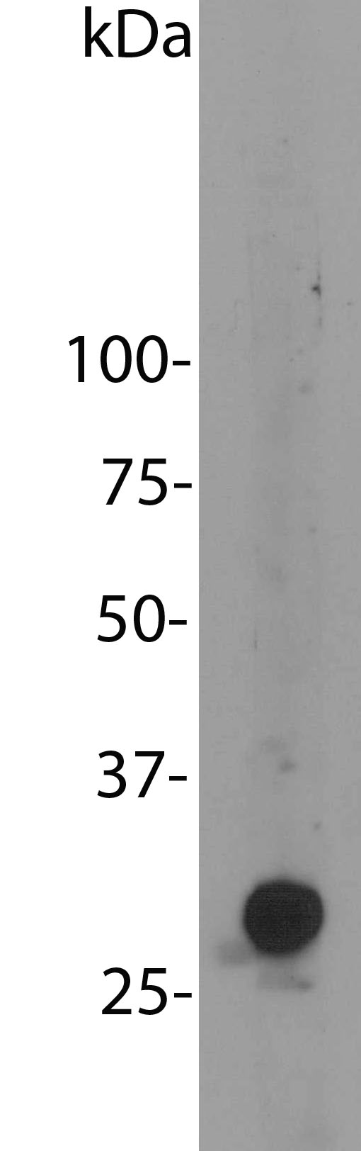
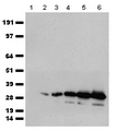
- Western blot (1:2000)
Usage
Try at dilutions of 1:1000 and higher for immunofluorescence. For western blots try at 1:2000. The mCherry protein runs at about 30kDa on SDS-PAGE gels.
Presentation
5 mM Sodium Azide
Storage
Store at 4°C.
Restrictions
For research use only. Intended for use by laboratory professionals.
LSBio Ratings
mCherry Antibody for ICC, IF/Immunofluorescence, WB/Western LS-C204825 has an LSBio Rating of
Publications (4.2)
Learn more about The LSBio Ratings Algorithm
Publications (3)
Chemogenetic Activation of an Extinction Neural Circuit Reduces Cue-Induced Reinstatement of Cocaine Seeking. Augur IF, Wyckoff AR, Aston-Jones G, Kalivas PW, Peters J. The Journal of neuroscience : the official journal of the Society for Neuroscience. 2016 36:10174-80. (IHC; Rat)
Selection and Characterization of Rupintrivir-Resistant Norwalk Virus Replicon Cells In Vitro. Kitano M, Hosmillo M, Emmott E, Lu J, Goodfellow I. Antimicrobial agents and chemotherapy. 2018 May;62:e00201-18. (Discosoma)
Behavioral and slice electrophysiological assessment of DREADD ligand, deschloroclozapine (DCZ) in rats. Todd B Nentwig , J Daniel Obray , Dylan T Vaughan, L Judson Chandler. Scientific reports. 2022 April;12:6595.
Customer Reviews (0)
Featured Products
Species:
Discosoma
Applications:
IHC, IHC - Paraffin, IHC - Frozen, Immunofluorescence, Western blot
Species:
Discosoma
Applications:
ICC, Immunofluorescence, Western blot
Source:
Human
Tag:
Myc-DDK (Flag)
Reactivity:
Porcine
Range:
0.31-20 ng/ml
Request SDS/MSDS
To request an SDS/MSDS form for this product, please contact our Technical Support department at:
Technical.Support@LSBio.com
Requested From: United States
Date Requested: 4/25/2024
Date Requested: 4/25/2024


