Login
Registration enables users to use special features of this website, such as past
order histories, retained contact details for faster checkout, review submissions, and special promotions.
order histories, retained contact details for faster checkout, review submissions, and special promotions.
Forgot password?
Registration enables users to use special features of this website, such as past
order histories, retained contact details for faster checkout, review submissions, and special promotions.
order histories, retained contact details for faster checkout, review submissions, and special promotions.
Quick Order
Products
Antibodies
ELISA and Assay Kits
Research Areas
Infectious Disease
Resources
Purchasing
Reference Material
Contact Us
Locations
Orders Processing,
Shipping & Receiving,
Warehouse
2 Shaker Rd Suites
B001/B101
Shirley, MA 01464
Production Lab
Floor 6, Suite 620
20700 44th Avenue W
Lynnwood, WA 98036
Telephone Numbers
Tel: +1 (206) 374-1102
Fax: +1 (206) 577-4565
Contact Us
Additional Contact Details
Login
Registration enables users to use special features of this website, such as past
order histories, retained contact details for faster checkout, review submissions, and special promotions.
order histories, retained contact details for faster checkout, review submissions, and special promotions.
Forgot password?
Registration enables users to use special features of this website, such as past
order histories, retained contact details for faster checkout, review submissions, and special promotions.
order histories, retained contact details for faster checkout, review submissions, and special promotions.
Quick Order
| Catalog Number | Size | Price |
|---|---|---|
| LS-C783540-100 | 100 µg (0.5 mg/ml) | $285 |
1 of 4
2 of 4
3 of 4
4 of 4
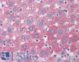
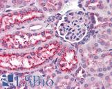
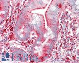
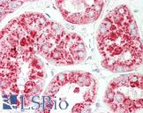
Monoclonal Mouse anti‑Human LAMP1 / CD107a Antibody (clone H4A3, IHC, WB) LS‑C783540
Monoclonal Mouse anti‑Human LAMP1 / CD107a Antibody (clone H4A3, IHC, WB) LS‑C783540
Antibody:
LAMP1 / CD107a Mouse anti-Human Monoclonal (H4A3) Antibody
Application:
IHC-P, ICC, WB, IP, Flo
Reactivity:
Human, Chimpanzee, Baboon, African green monkey, Cynomolgus monkey, Pig-tailed macaque, Rhesus monkey
Format:
Unconjugated, Unmodified
Toll Free North America
 206-374-1102
For Research Use Only
Overview
Antibody:
LAMP1 / CD107a Mouse anti-Human Monoclonal (H4A3) Antibody
Application:
IHC-P, ICC, WB, IP, Flo
Reactivity:
Human, Chimpanzee, Baboon, African green monkey, Cynomolgus monkey, Pig-tailed macaque, Rhesus monkey
Format:
Unconjugated, Unmodified
Specifications
Description
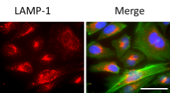
CD107a antibody LS-C783540 is an unconjugated mouse monoclonal antibody to CD107a (LAMP1) from human. It is reactive with human, african green monkey, baboon and other species. Validated for Flow, ICC, IHC, IP and WB.
Target
Human LAMP1 / CD107a
Synonyms
LAMP1 | CD107a | CD107a antigen | LAMP-1 | LGP120 | LAMPA
Host
Mouse
Reactivity
Human, Chimpanzee, Baboon, African green monkey, Cynomolgus monkey, Pig-tailed macaque, Rhesus monkey
(tested or 100% immunogen sequence identity)
Clonality
IgG1,k
Monoclonal
Clone
H4A3
Conjugations
Unconjugated
Purification
Purified by affinity chromatography.
Modifications
Unmodified
Immunogen
Human adult adherent peripheral blood cells
Applications
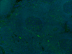
- IHC - Paraffin (5 - 10 µg/ml)
- ICC
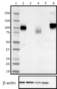
- Western blot (1 - 5 µg/ml)
- Immunoprecipitation
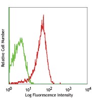
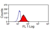
- Flow Cytometry (2 µg/10E6 cells)

|
Performing IHC? See our complete line of Immunohistochemistry Reagents including antigen retrieval solutions, blocking agents
ABC Detection Kits and polymers, biotinylated secondary antibodies, substrates and more.
|
Usage
Each lot of this antibody is quality control tested by immunofluorescent staining with flow cytometric analysis. For flow cytometric staining, the suggested use of this reagent is = 2.0 µg per million cells in 100 µl volume. For Western blotting, the suggested use of this reagent is 1.0 - 5.0 µg per ml. For immunocytochemistry on formalin-fixed paraffin-embedded tissue sections, a concentration range of 5.0 - 10 µg per ml is recommended. It is recommended that the reagent be titrated for optimal performance for each application. Additional reported applications (for the relevant formats) include: Western blotting, immunohistochemical staining, immunofluorescence, and immunoprecipitation.This antibody is specific to human LAMP-1. Positive control: Hela cells; LAMP-1 molecular weight appears to be at ~110 kDa on the gel due to high glycosylation.
Presentation
PBS pH 7.2, 0.09% sodium azide.
Storage
Store at 2°C to 8°C.
Restrictions
For research use only. Intended for use by laboratory professionals.
About LAMP1 / CD107a
Publications (0)
Customer Reviews (0)
Featured Products
Species:
Human, Mouse, Rat
Applications:
IHC, IHC - Paraffin, Immunofluorescence, Western blot, ELISA
Species:
Mouse
Applications:
IHC, IHC - Paraffin, IHC - Frozen, Western blot, Immunoprecipitation, Flow Cytometry
Species:
Human
Applications:
IHC, IHC - Paraffin, ICC, Immunofluorescence, Western blot
Species:
Human, Mouse, Non-Human Primates
Applications:
IHC, IHC - Paraffin, IHC - Frozen, ICC, Western blot, Flow Cytometry
Species:
Pig
Applications:
IHC, IHC - Paraffin, IHC - Frozen, Western blot, Immunoprecipitation, Flow Cytometry
Request SDS/MSDS
To request an SDS/MSDS form for this product, please contact our Technical Support department at:
Technical.Support@LSBio.com
Requested From: United States
Date Requested: 4/18/2024
Date Requested: 4/18/2024


