Login
Registration enables users to use special features of this website, such as past
order histories, retained contact details for faster checkout, review submissions, and special promotions.
order histories, retained contact details for faster checkout, review submissions, and special promotions.
Forgot password?
Registration enables users to use special features of this website, such as past
order histories, retained contact details for faster checkout, review submissions, and special promotions.
order histories, retained contact details for faster checkout, review submissions, and special promotions.
Quick Order
Products
Antibodies
ELISA and Assay Kits
Research Areas
Infectious Disease
Resources
Purchasing
Reference Material
Contact Us
Locations
Orders Processing,
Shipping & Receiving,
Warehouse
2 Shaker Rd Suites
B001/B101
Shirley, MA 01464
Production Lab
Floor 6, Suite 620
20700 44th Avenue W
Lynnwood, WA 98036
Telephone Numbers
Tel: +1 (206) 374-1102
Fax: +1 (206) 577-4565
Contact Us
Additional Contact Details
Login
Registration enables users to use special features of this website, such as past
order histories, retained contact details for faster checkout, review submissions, and special promotions.
order histories, retained contact details for faster checkout, review submissions, and special promotions.
Forgot password?
Registration enables users to use special features of this website, such as past
order histories, retained contact details for faster checkout, review submissions, and special promotions.
order histories, retained contact details for faster checkout, review submissions, and special promotions.
Quick Order
| Catalog Number | Size | Price |
|---|---|---|
| LS-C55014-100 | 100 µg (0.5 mg/ml) | $503 |
1 of 5
2 of 5
3 of 5
4 of 5
5 of 5
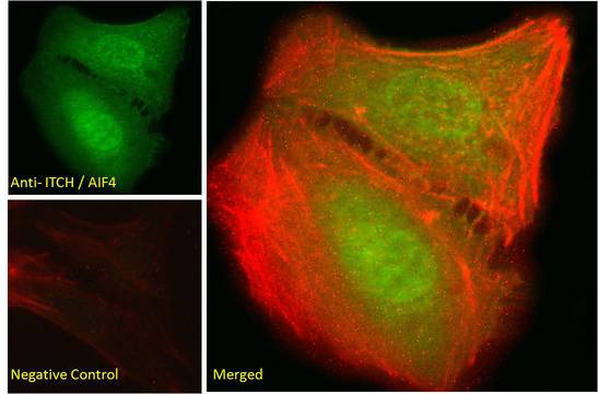
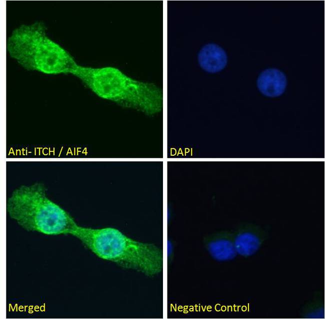
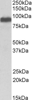
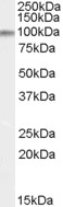
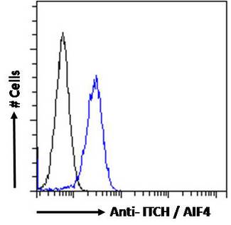
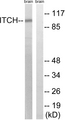
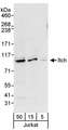
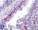
Polyclonal Goat anti‑Human ITCH / AIP4 Antibody (aa714‑726, IF, WB) LS‑C55014
Polyclonal Goat anti‑Human ITCH / AIP4 Antibody (aa714‑726, IF, WB) LS‑C55014
Antibody:
ITCH / AIP4 Goat anti-Human Polyclonal (aa714-726) Antibody
Application:
IF, WB, Peptide-ELISA
Reactivity:
Human, Monkey, Mouse, Rat, Bat, Bovine, Dog, Hamster, Horse, Pig, Rabbit
Format:
Unconjugated, Unmodified
Toll Free North America
 206-374-1102
For Research Use Only
Overview
Antibody:
ITCH / AIP4 Goat anti-Human Polyclonal (aa714-726) Antibody
Application:
IF, WB, Peptide-ELISA
Reactivity:
Human, Monkey, Mouse, Rat, Bat, Bovine, Dog, Hamster, Horse, Pig, Rabbit
Format:
Unconjugated, Unmodified
Specifications
Description
AIP4 antibody LS-C55014 is an unconjugated goat polyclonal antibody to AIP4 (ITCH) (aa714-726) from human. It is reactive with human, mouse, rat and other species. Validated for IF, Peptide-ELISA and WB.
Target
Human ITCH / AIP4
Synonyms
ITCH | AIF4 | AIP4 | DJ468O1.1 | NAPP1 | NFE2-associated polypeptide 1
Host
Goat
Reactivity
Human, Monkey, Mouse, Rat, Bat, Bovine, Dog, Hamster, Horse, Pig, Rabbit
(tested or 100% immunogen sequence identity)
Clonality
Polyclonal
Conjugations
Unconjugated
Purification
Purified from goat serum by ammonium sulphate precipitation followed by antigen affinity chromatography using the immunizing peptide.
Modifications
Unmodified
Immunogen
Peptide with sequence C-EIKSHDLKPNGGN, from the internal region of the protein sequence according to NP_113671.3NP_001244066.1NP_001244067.1.
Epitope
aa714-726
Specificity
Human ITCH.
Applications
- Immunofluorescence (10 µg/ml)
- Western blot (0.1 - 0.3 µg/ml)
- Peptide Enzyme-Linked Immunosorbent Assay (1:16000)
Presentation
TBS, pH 7.3, 0.02% Sodium Azide, 0.5% BSA
Storage
Aliquot and store at -20°C. Avoid freeze-thaw cycles.
Restrictions
For research use only. Intended for use by laboratory professionals.
About ITCH / AIP4
Publications (0)
Customer Reviews (0)
Featured Products
Species:
Human, Mouse
Applications:
Western blot, Peptide Enzyme-Linked Immunosorbent Assay
Reactivity:
Human, Mouse
Range:
Positive/Negative
Species:
Human
Applications:
IHC, IHC - Paraffin, Immunofluorescence, Western blot, ELISA
Request SDS/MSDS
To request an SDS/MSDS form for this product, please contact our Technical Support department at:
Technical.Support@LSBio.com
Requested From: United States
Date Requested: 4/17/2024
Date Requested: 4/17/2024


