Login
Registration enables users to use special features of this website, such as past
order histories, retained contact details for faster checkout, review submissions, and special promotions.
order histories, retained contact details for faster checkout, review submissions, and special promotions.
Forgot password?
Registration enables users to use special features of this website, such as past
order histories, retained contact details for faster checkout, review submissions, and special promotions.
order histories, retained contact details for faster checkout, review submissions, and special promotions.
Quick Order
Products
Antibodies
ELISA and Assay Kits
Research Areas
Infectious Disease
Resources
Purchasing
Reference Material
Contact Us
Locations
Orders Processing,
Shipping & Receiving,
Warehouse
2 Shaker Rd Suites
B001/B101
Shirley, MA 01464
Production Lab
Floor 6, Suite 620
20700 44th Avenue W
Lynnwood, WA 98036
Telephone Numbers
Tel: +1 (206) 374-1102
Fax: +1 (206) 577-4565
Contact Us
Additional Contact Details
Login
Registration enables users to use special features of this website, such as past
order histories, retained contact details for faster checkout, review submissions, and special promotions.
order histories, retained contact details for faster checkout, review submissions, and special promotions.
Forgot password?
Registration enables users to use special features of this website, such as past
order histories, retained contact details for faster checkout, review submissions, and special promotions.
order histories, retained contact details for faster checkout, review submissions, and special promotions.
Quick Order
| Catalog Number | Size | Price |
|---|---|---|
| LS-C18827-1 | 1 mg (2 mg/ml) | $1,225 |
1 of 2
2 of 2
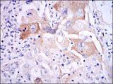
Polyclonal Rabbit anti‑Human IL‑1B / IL‑1 Beta Antibody (N‑Terminus, IHC, IF, WB) LS‑C18827
Polyclonal Rabbit anti‑Human IL‑1B / IL‑1 Beta Antibody (N‑Terminus, IHC, IF, WB) LS‑C18827

Note: This antibody replaces LS-C7719, LS-C70852
Antibody:
IL-1B / IL-1 Beta Rabbit anti-Human Polyclonal (N-Terminus) Antibody
Application:
IHC, IHC-P, IHC-Fr, IF, WB, IP, Flo, ELISA, Neut, RIA
Reactivity:
Human, Dog, Primate
Format:
Unconjugated, Unmodified
Toll Free North America
 206-374-1102
For Research Use Only
Overview
Antibody:
IL-1B / IL-1 Beta Rabbit anti-Human Polyclonal (N-Terminus) Antibody
Application:
IHC, IHC-P, IHC-Fr, IF, WB, IP, Flo, ELISA, Neut, RIA
Reactivity:
Human, Dog, Primate
Format:
Unconjugated, Unmodified
Specifications
Description
IL-1 Beta antibody LS-C18827 is an unconjugated rabbit polyclonal antibody to IL-1 Beta (IL-1B) (N-Terminus) from human. It is reactive with human, dog and primate. Validated for ELISA, Flow, IF, IHC, IP, Neut, RIA and WB. Cited in 2 publications.
Target
Human IL-1B / IL-1 Beta
Synonyms
IL1B | IL-1B | IL-1 beta | IL1-BETA | Preinterleukin 1 beta | Pro-interleukin-1-beta | Catabolin | IL-1 | IL1F2 | Interleukin 1, beta | Interleukin-1 beta
Host
Rabbit
Reactivity
Human, Dog, Primate
(tested or 100% immunogen sequence identity)
Clonality
IgG
Polyclonal
Conjugations
Unconjugated
Purification
DEAE fractionation
Modifications
Unmodified
Immunogen
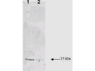
This antibody was prepared by repeated immunizations with recombinant human IL-1ß produced in E.coli. The MW of the recombinant 153 aa IL-1ß was 17 kDa with the N-terminal amino acid at position alanine 117. This cleavage site is generated by the IL-1ß converting enzyme (ICE, capase-1).
Epitope
N-Terminus
Specificity
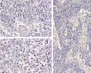
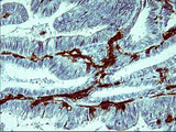
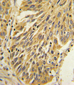
This antibody is primarily directed against mature, 17,000 MW human IL-1ß and is useful in determining its presence in various assays. In general, this antibody also detects primate IL-1ß in the same formats using similar dilutions. The antiserum does not recognize human IL-1a. In ELISA formats and other immunoreactive assays, this antibody will recognize 10% of the non-denatured (native) precursor 31,000 MW IL-1ß containing samples but will primarily detect all of the 17,000 MW mature molecule. However, in immunoblot analysis of natural cell products or human body fluids, the usual procedure of hearing the sample in SDS with or without reducing agents will facilitate denaturing of the 31,000 MW IL- 1ß precursor molecule. Denatured 31,000 precursor IL-1ß will be recognized by this antibody but often migrates as a 35,000 MW band. This is due to the unfolding of the denatured precursor IL-1ß exposing epitopes not exposed in the natural state. In immunoblots, depending on the number of cells, the antibody detects the 17,000 MW band in supernatants as well as a 35,000 MW band representing the 31,000 MW IL-1ß precursor in lysates.
Applications
- IHC
- IHC - Paraffin (1:100 - 1:200)
- IHC - Frozen (1:100 - 1:200)
- Immunofluorescence
- Western blot (1:1000)
- Immunoprecipitation (1:400 - 1:800)
- Flow Cytometry
- ELISA (1:500 - 1:2000)
- Neutralization (1:100)
- Radioimmunoassay (1:5000 - 1:10000)

|
Performing IHC? See our complete line of Immunohistochemistry Reagents including antigen retrieval solutions, blocking agents
ABC Detection Kits and polymers, biotinylated secondary antibodies, substrates and more.
|
Usage
This IgG fraction antibody of anti- Human IL-1beta has been tested for use in neutralizations, ELISA, radioimmunoassays, immunohistochemistry, immunoblotting and immunoprecipitation. It recognizes the 17000 MW mature IL-10. For immunoblots, typically, IL-1beta is detected from supernatants or lysates of 2 x 10⁶ endotoxin-stimulated human peripheral blood mononuclear cells (PBMC). PBMC are stimulated for 24 hours with 1 % (v/v) human serum plus 10 ng/mL E. coli LPS. For immunoprecipitation a dilution of 1:400 to 1:800 is suggested. Pre-clearing the preparation with a non-specific rabbit IgG (011-001-297) to reduce background. For immunohistochemistry a dilution of 1:100 to 1:200 is suggested. Either paraffin fixation or cryofixation can be used for sample preparation to stain intracellular IL- 1beta. For ELISA also use HRP Conjugated Anti-Rabbit IgG [HalphaL] (Goat) (611-1302) for detection.
Presentation
0.02 M Potassium Phosphate, pH 7.2, 0.15 M NaCl
Storage
Aliquot and store at -20°C. Avoid freeze-thaw cycles.
Restrictions
For research use only. Intended for use by laboratory professionals.
About IL-1B / IL-1 Beta
LSBio Ratings
IL-1B / IL-1 Beta Antibody (N-Terminus) for IHC, IF/Immunofluorescence, WB/Western, IP, Flow, ELISA LS-C18827 has an LSBio Rating of
Publications (4.1)
Learn more about The LSBio Ratings Algorithm
Publications (2)
Serum soluble interleukin 7 receptor is strongly associated with lupus nephritis in patients with systemic lupus erythematosus. Badot V, Luijten RK, van Roon JA, Depresseux G, Aydin S, Van den Eynde BJ, Houssiau FA, Lauwerys BR. Annals of the rheumatic diseases. 2013 72:453-6. (IHC-Fr; Human)
p65-Dependent production of interleukin-1 beta by osteolytic prostate cancer cells causes an induction of chemokine expression in osteoblasts. Schulze J, Weber K, Baranowsky A, Streichert T, Lange T, Spiro AS, Albers J, Seitz S, Zustin J, Amling M, Fehse B, Schinke T. Cancer letters. 2012 Apr;317:106-13.
Customer Reviews (0)
Featured Products
Species:
Human
Applications:
IHC, IHC - Paraffin, Immunofluorescence, Western blot
Species:
Human
Applications:
IHC, IHC - Paraffin, Western blot, Flow Cytometry
Species:
Human
Applications:
IHC, IHC - Paraffin, Immunofluorescence, Western blot, ELISA
Reactivity:
Chicken
Range:
7.8-500 pg/ml
Request SDS/MSDS
To request an SDS/MSDS form for this product, please contact our Technical Support department at:
Technical.Support@LSBio.com
Requested From: United States
Date Requested: 4/16/2024
Date Requested: 4/16/2024


