Login
Registration enables users to use special features of this website, such as past
order histories, retained contact details for faster checkout, review submissions, and special promotions.
order histories, retained contact details for faster checkout, review submissions, and special promotions.
Forgot password?
Registration enables users to use special features of this website, such as past
order histories, retained contact details for faster checkout, review submissions, and special promotions.
order histories, retained contact details for faster checkout, review submissions, and special promotions.
Quick Order
Products
Antibodies
ELISA and Assay Kits
Research Areas
Infectious Disease
Resources
Purchasing
Reference Material
Contact Us
Locations
Orders Processing,
Shipping & Receiving,
Warehouse
2 Shaker Rd Suites
B001/B101
Shirley, MA 01464
Production Lab
Floor 6, Suite 620
20700 44th Avenue W
Lynnwood, WA 98036
Telephone Numbers
Tel: +1 (206) 374-1102
Fax: +1 (206) 577-4565
Contact Us
Additional Contact Details
Login
Registration enables users to use special features of this website, such as past
order histories, retained contact details for faster checkout, review submissions, and special promotions.
order histories, retained contact details for faster checkout, review submissions, and special promotions.
Forgot password?
Registration enables users to use special features of this website, such as past
order histories, retained contact details for faster checkout, review submissions, and special promotions.
order histories, retained contact details for faster checkout, review submissions, and special promotions.
Quick Order
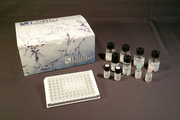
| Catalog Number | Size | Price |
|---|---|---|
| LS-C153837-100 | 100 µg (1 mg/ml) | $574 |
1 of 4
2 of 4
3 of 4
4 of 4
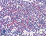
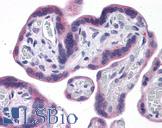
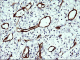
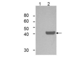
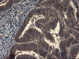
Monoclonal Mouse anti‑Mouse IDO1 / IDO Antibody (clone 2E2.6, IHC, IF, WB) LS‑C153837
Monoclonal Mouse anti‑Mouse IDO1 / IDO Antibody (clone 2E2.6, IHC, IF, WB) LS‑C153837

Antibody:
IDO1 / IDO Mouse anti-Mouse Monoclonal (2E2.6) Antibody
Application:
IHC, ICC, IF, WB, Flo, ELISA
Reactivity:
Mouse
Format:
Unconjugated, Unmodified
Toll Free North America
 206-374-1102
For Research Use Only
Overview
Antibody:
IDO1 / IDO Mouse anti-Mouse Monoclonal (2E2.6) Antibody
Application:
IHC, ICC, IF, WB, Flo, ELISA
Reactivity:
Mouse
Format:
Unconjugated, Unmodified
Specifications
Description
IDO antibody LS-C153837 is an unconjugated mouse monoclonal antibody to mouse IDO (IDO1). Validated for ELISA, Flow, ICC, IF, IHC and WB. Cited in 1 publication.
Target
Mouse IDO1 / IDO
Synonyms
IDO1 | IDO | Indole 2,3-dioxygenase | Indolamine 2,3 dioxygenase | IDO-1 | INDO | Indoleamine 2,3-dioxygenase | Indoleamine 2,3-dioxygenase 1
Host
Mouse
Reactivity
Mouse
(tested or 100% immunogen sequence identity)
Non-Reactivity
Human
Clonality
IgG1
Monoclonal
Clone
2E2.6
Conjugations
Unconjugated
Purification
Protein A affinity chromatography
Modifications
Unmodified
Immunogen
IDO1 antibody was produced in mouse by repeated immunizations with mouse recombinant IDO1 protein followed by hybridoma development.
Specificity
Mouse IDO1 does not react with human tissues. Cross-reactivity with IDO1 from other sources has not been determined.
Applications
- IHC
- ICC
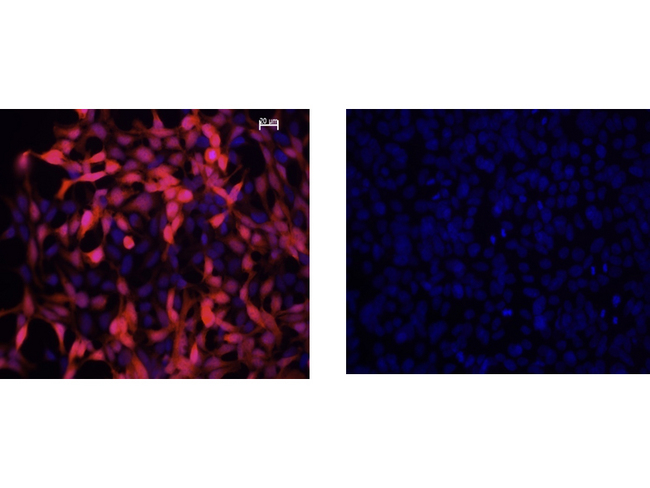
- Immunofluorescence (1:50 - 1:100)
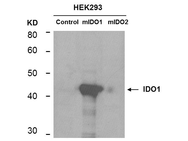
- Western blot (1:500 - 1:1500)
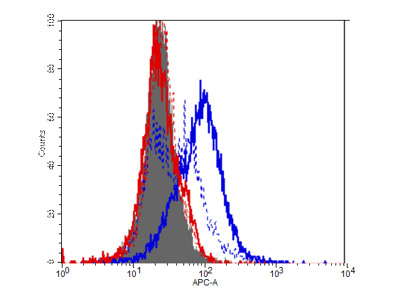
- Flow Cytometry
- ELISA (1:5000 - 1:50000)

|
Performing IHC? See our complete line of Immunohistochemistry Reagents including antigen retrieval solutions, blocking agents
ABC Detection Kits and polymers, biotinylated secondary antibodies, substrates and more.
|
Usage
Anti-IDO1 antibody has been tested for use in ELISA, Western Blot, IF, IHC, and Flow Cytometry. Specific conditions for reactivity should be optimized by the end user.
Presentation
0.02 M Potassium Phosphate, pH 7.2, 0.15 M NaCl, 0.01% Sodium Azide
Storage
Short term: store at 4°C. Long term: aliquot and store at -20°C. Avoid freeze-thaw cycles.
Restrictions
For research use only. Intended for use by laboratory professionals.
About IDO1 / IDO
LSBio Ratings
IDO1 / IDO Antibody (clone 2E2.6) for IHC, ICC, IF/Immunofluorescence, WB/Western, Flow, ELISA LS-C153837 has an LSBio Rating of
Publications (4)
Learn more about The LSBio Ratings Algorithm
Publications (1)
Regulation of indoleamine 2, 3-dioxygenase in hippocampal microglia by NLRP3 inflammasome in lipopolysaccharide-induced depressive-like behaviors. Shanshan Zhang, Ying Zong, Zhonggan Ren, Juntao Hu, Xinyuan Wu, Honglei Xiao, Song Qin, Guomin Zhou, Yuanyuan Ma, Yaodong Zhang, Jin Yu, Kaidi Wang, Guocai Lu, Qiong Liu. The European journal of neuroscience. 2020 December;52:4586-4601.
Customer Reviews (0)
Featured Products
Species:
Human, Monkey, Mouse
Applications:
IHC, IHC - Paraffin, Western blot, Peptide Enzyme-Linked Immunosorbent Assay
Species:
Human
Applications:
IHC, IHC - Paraffin, Western blot, ELISA
Species:
Human
Applications:
IHC, IHC - Paraffin, Immunofluorescence, Western blot
Species:
Human, Mouse
Applications:
IHC, Immunofluorescence, Western blot, Immunoprecipitation, ELISA
Species:
Human
Applications:
IHC, IHC - Paraffin, Immunofluorescence, Western blot
Request SDS/MSDS
To request an SDS/MSDS form for this product, please contact our Technical Support department at:
Technical.Support@LSBio.com
Requested From: United States
Date Requested: 4/16/2024
Date Requested: 4/16/2024


